News

Researchers at the MPI of Biochemistry, together with collaborating partners, have discovered the first proteomic subtype of an aggressive blood cancer known as acute myeloid leukemia by using mass spectrometry technology.

In order to better treat patients diagnosed with acute myeloid leukemia (AML), the pathological processes and also existing subtypes of the disease must be better understood. With the help of proteome and genome analysis, researchers at the Max Planck Institute (MPI) of Biochemistry in Martinsried, together with cooperation partners from the University Hospital in Frankfurt am Main, have discovered a new subtype. This subgroup contains elevated levels of mitochondrial proteins and thus has altered mitochondrial metabolism. These so-called mito-AML cells can be combated more effectively in laboratory experiments with the help of inhibitors against mitochondrial respiration than with conventional chemotherapeutic agents. The study was published in Cancer Cell.

Identification of molecular AML subtypes

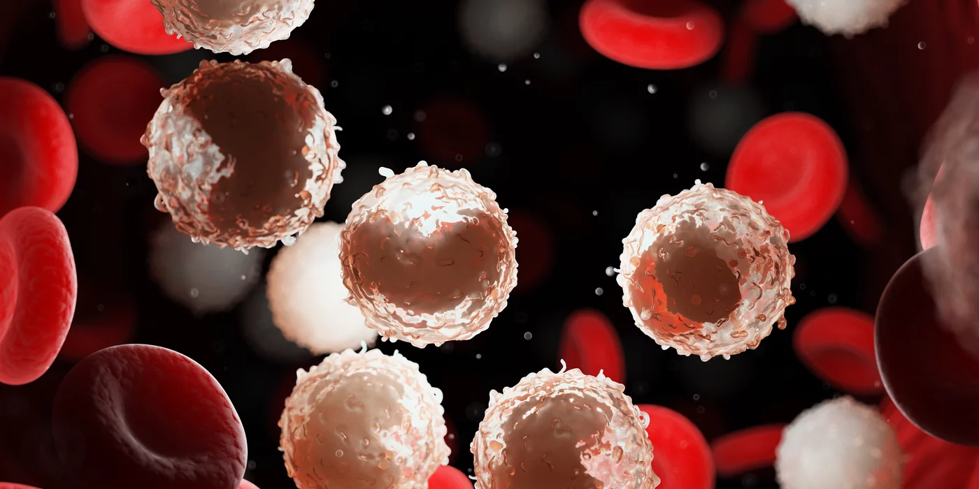
Acute myeloid leukemia (AML) is an aggressive cancer originating from blood cells. When immature blood cells in the bone marrow acquire certain aberrations in their genome they become malignant and overgrow the bone marrow, the place where normally blood cells are produced. As a consequence, normal blood cells are suppressed by the leukemia cells and this leads to infections, bleeding and ultimately death of patients. Most patients diagnosed with AML undergo chemotherapy. In the last decades genomic studies identified molecular subtypes of the disease thereby opening up a perspective for personalized therapeutic approaches in AML. As a result, Clinicians and researchers now distinguish different genomic AML subtypes and for some of them they now even use specific therapeutics. These discoveries have certainly revolutionized the molecular understanding of the disease. However, despite this progress, prognosis for AML remains poor, highlighting the strong medical need for a deeper understanding of AML pathophysiology and for further innovative and more efficient therapies. 

Read more


graduationCongratulations on your PhD!

Mai Ly Tran


Mechanisms of secretory cargo sorting at the trans-Golgi network  

RG: Julia von Blume


Publication Placeholder

Scherr, M.J., Wahab, S.A., Remus, D., and Duderstadt, K.E.
Cell Rep, 2022, 38, 110531.
doi: 10.1016/j.celrep.2022.110531

Mobile origin-licensing factors confer resistance to conflicts with RNA polymerase

Fundamental to our understanding of chromosome duplication is the idea that replication origins function both as sites where MCM helicases are loaded during the G1 phase and where synthesis begins in S phase. However, the temporal delay between phases exposes the replisome assembly pathway to potential disruption prior to replication. Using multicolor, single-molecule imaging, we systematically study the consequences of encounters between actively transcribing RNA polymerases (RNAPs) and replication initiation intermediates in the context of chromatin. We demonstrate that RNAP can push multiple licensed MCM helicases over long distances with nucleosomes ejected or displaced. Unexpectedly, we observe that MCM helicase loading intermediates also can be repositioned by RNAP and continue origin licensing after encounters with RNAP, providing a web of alternative origin specification pathways. Taken together, our observations reveal a surprising mobility in origin-licensing factors that confers resistance to the complex challenges posed by diverse obstacles encountered on chromosomes.


Publication Placeholder

Weickert, P., and Stingele, J.
Annu Rev Biochem, 2022, online ahead of print.
doi: 10.1146/annurev-biochem-032620-105820

DNA-Protein Crosslinks and Their Resolution

Covalent DNA-protein crosslinks (DPCs) are pervasive DNA lesions that interfere with essential chromatin processes such as transcription or replication. This review strives to provide an overview of the sources and principles of cellular DPC formation. DPCs are caused by endogenous reactive metabolites and various chemotherapeutic agents. However, in certain conditions DPCs also arise physiologically in cells. We discuss the cellular mechanisms resolving these threats to genomic integrity. Detection and repair of DPCs require not only the action of canonical DNA repair pathways but also the activity of specialized proteolytic enzymes-including proteases of the SPRTN/Wss1 family-to degrade the crosslinked protein. Loss of DPC repair capacity has dramatic consequences, ranging from genome instability in yeast and worms to cancer predisposition and premature aging in mice and humans.


Publication Placeholder

Unterauer, E.M., and Jungmann, R.
Front Synaptic Neurosci, 2022, 13, 798267.
doi: 10.3389/fnsyn.2021.798267

Quantitative Imaging With DNA-PAINT for Applications in Synaptic Neuroscience

Super-resolution (SR) microscopy techniques have been advancing the understanding of neuronal protein networks and interactions. Unraveling the arrangement of proteins with molecular resolution provided novel insights into neuron cytoskeleton structure and actin polymerization dynamics in synaptic spines. Recent improvements in quantitative SR imaging have been applied to synaptic protein clusters and with improved multiplexing technology, the interplay of multiple protein partners in synaptic active zones has been elucidated. While all SR techniques come with benefits and drawbacks, true molecular quantification is a major challenge with the most complex requirements for labeling reagents and careful experimental design. In this perspective, we provide an overview of quantitative SR multiplexing and discuss in greater detail the quantification and multiplexing capabilities of the SR technique DNA-PAINT. Using predictable binding kinetics of short oligonucleotides, DNA-PAINT provides two unique approaches to address multiplexed molecular quantification: qPAINT and Exchange-PAINT. With precise and accurate quantification and spectrally unlimited multiplexing, DNA-PAINT offers an attractive route to unravel complex protein interaction networks in neurons. Finally, while the SR community has been pushing technological advances from an imaging technique perspective, the development of universally available, small, efficient, and quantitative labels remains a major challenge in the field.


graduationCongratulations on your PhD!

Diana Inês Lopes Amaro


Neuronal Representation of Sound Source Location in the Auditory Cortex during Active Navigation 

RG: Benedikt Grothe


graduationCongratulations on your PhD!

Jan Rieckmann


Immune Cell Proteomes 

RG: Felix Meißner


Publication Placeholder

Brandstetter, K., Zülske, T., Ragoczy, T., Hörl, D., Guirao-Ortiz, M., Steinek, C., Barnes, T., Stumberger, G., Schwach, J., Haugen, E., Rynes, E., Korber, P., Stamatoyannopoulos, J.A., Leonhardt, H., Wedemann, G., and Harz, H.
(IMPRS-LS students are in bold)
Biophys J., 2022, online ahead of print.
doi: 10.1016/j.bpj.2022.02.009

Differences in nanoscale organization of regulatory active and inactive human chromatin

Methodological advances in conformation capture techniques have fundamentally changed our understanding of chromatin architecture. However, the nanoscale organization of chromatin and its cell-to-cell variance are less studied. Analyzing genome-wide data from 733 human cell and tissue samples, we identified 2 prototypical regions that exhibit high or absent hypersensitivity to deoxyribonuclease I, respectively. These regulatory active or inactive regions were examined in the lymphoblast cell line K562 by using high-throughput super-resolution microscopy. In both regions, we systematically measured the physical distance of 2 fluorescence in situ hybridization spots spaced by only 5 kb of DNA. Unexpectedly, the resulting distance distributions range from very compact to almost elongated configurations of more than 200-nm length for both the active and inactive regions. Monte Carlo simulations of a coarse-grained model of these chromatin regions based on published data of nucleosome occupancy in K562 cells were performed to understand the underlying mechanisms. There was no parameter set for the simulation model that can explain the microscopically measured distance distributions. Obviously, the chromatin state given by the strength of internucleosomal interaction, nucleosome occupancy, or amount of histone H1 differs from cell to cell, which results in the observed broad distance distributions. This large variability was not expected, especially in inactive regions. The results for the mechanisms for different distance distributions on this scale are important for understanding the contacts that mediate gene regulation. Microscopic measurements show that the inactive region investigated here is expected to be embedded in a more compact chromatin environment. The simulation results of this region require an increase in the strength of internucleosomal interactions. It may be speculated that the higher density of chromatin is caused by the increased internucleosomal interaction strength.


A team from the Max Planck Institue of Biochemistry in Martinsried and the Martin Luther University Halle-Wittenberg has discovered how an essential final step in the production of mRNA precisely works.

Proteins need to interact in a complex manner for a so-called “messenger RNA” (mRNA) to be created in human cells from a precursor molecule. mRNA provides a blueprint for proteins; the first vaccines against the coronavirus are also based on mRNAs. A team from the Max Planck Institute (MPI) of Biochemistry in Martinsried and the Martin Luther University Halle-Wittenberg (MLU) has discovered how an essential final step in the production of mRNA precisely works. The study was published in Genes & Development.

Proteins are responsible for all of the body’s essential processes. In a sense, the genes in the human genome act as building instructions for them. However, an intermediate step is necessary before new proteins can be created: “First the DNA must be transcribed: A chain-like precursor RNA is produced which is an exact copy of the DNA. From this, several steps are required to create the mature mRNA. This process is essential for the cell to build new proteins,” says biochemist Professor Elmar Wahle from MLU who led the team alongside Professor Elena Conti, an expert in structural biology at the MPI of Biochemistry. 

Read more


graduationCongratulations on your PhD!

Matthias Scherr


The Dynamics of Eukaryotic Replication Initiation Revealed at the Single-Molecule Level 

RG: Karl Duderstadt