Functional ultrasound imaging of brain-wide activity
A team of scientists from the Neuro-Electronics Research Flanders developed and tested, in cooperation with scientists from the MPI of Neurobiology, a new volumetric functional ultrasound imaging platform. The ease of use, reliability, and affordability of the technology make it an excellent candidate for driving future brain-wide neuroimaging research.
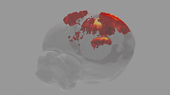
Ultrasound is used to image soft tissue or organs, such as the heart, lungs and bladder, in real time. It is routinely used in hospitals because it is both safe and affordable. Over the past ten years, scientists at the Urban Lab (NERF, empowered by imec, KU Leuven and VIB) have contributed to the development of innovative brain ultrasound hardware and software solutions in collaboration with several academic and industrial partners. Using functional ultrasound imaging (fUSI) they have succeeded to visualize neural activity by mapping local changes in cerebral blood flow. Initially, however, fUSI was restricted to cross-sectional 2D imaging within a small field of view, which meant that visualization of brain-wide activity remained a challenge.