News

In December 2015, Facebook founder Mark Zuckerberg and his wife Priscilla Chan announced their plan to put 99% percent of their Facebook shares, worth about 45 billion dollars, into a new project focusing on the improvement of human potential and the promotion of equality. After an initial focus on education, the initiative has now announced its second focal point: the promotion of basic science research with the goal of curing, preventing or managing all disease by the end of this century. This new project, led by neuroscientist Cori Bargmann, was announced in San Francisco yesterday. In addition to the President, the two founders also introduced the scientists who will help build this initiative in the future. The panel of experts also includes one German researcher: Tobias Bonhoeffer, Director at the Max Planck Institute of Neubiology.

Shortly after the birth of their daughter Maxima Chan Zuckerberg, the couple set up the Chan Zuckerberg Initiative, stating they wished to see their daughter grow up in a world in which it was possible to cure diseases, personalize learning, and connect people. At a press conference on the Mission Bay campus of the University of California in San Francisco yesterday, Chan and Zuckerberg announced the second cornerstone of the initiative: 'Chan Zuckerberg Science'.

Over the past few months, the paediatrician Chan and Facebook founder Zuckerberg had met with numerous scientists, engineers, and other experts. This convinced them of the importance to invest into basic research to achieve much needed scientific progress essential for combating diseases. As a result, they decided to set up "Chan Zuckerberg Science (CZS)". The work of the initiative will have three core elements: More and better cooperation of scientists and engineers, the development of new methods and technologies, and educating the general public about the benefits of scientific discovery with the result of a much more general support and willingness to donate to science.

The only European member of the Scientific Advisory Board is Max Planck Director Tobias Bonhoeffer from Martinsried. "I am truly thrilled to be part of this exciting initiative. I am convinced that CZS can provide a powerful new impetus for imaginative and forward-looking science in the biomedical arena", was his first reaction. "It is a great pleasure and honor for me to have been appointed as advisor," said Bonhoeffer, who had been in contact with Chan and Zuckerberg since their visit to Berlin earlier this year. Being involved in major science policy decisions is however nothing new for the 56-year-old neurobiologist Tobias Bonhoeffer. In his capacity as chairman of the Biology and Medicine Section of the Max Planck Society (2008 - 2011) and as a Governor of the Wellcome Trust, one of the world's largest charities, he has provided guidance and expertise to many decision-making bodies.

Read more


Changes in gene activity are fundamental to many neuropsychiatric disorders. However, the nature of the genes affected, the time and place in which these changes occur, or the impact of these changes on nerve cells, remain largely unknown. One of the reasons for this ignorance is the inability to tag individual genes and their products without disrupting normal cell functions. An international team from the Max Planck Institute of Neurobiology, the University of Birmingham, the Wigner Institute in Budapest and Femtonics Ltd. now aim to close this gap in methodology. The European Union supports the project with a Horizon 2020 grant, worth more than four million Euros.

Many diseases also have a genetic component. For example, gene mutations can lead to the development of cancer or psychiatric disorders such as schizophrenia, depression or autism. Most of these diseases seem to be caused not by a single mutation but are rather the result of the interplay of dozens, maybe even hundreds of altered gene functions. It would therefore be of great benefit for the investigation, diagnosis and therapy of these illnesses, to identify the affected genes and monitor their activity in the brain of the living organism.
An interdisciplinary team around Herwig Baier from the Max Planck Institute of Neurobiology, Attila Sik from the University of Birmingham, Miklós Veres from the Wigner Institute and the microscope developers at Femtonics Ltd. aim to develop such a method. (more)


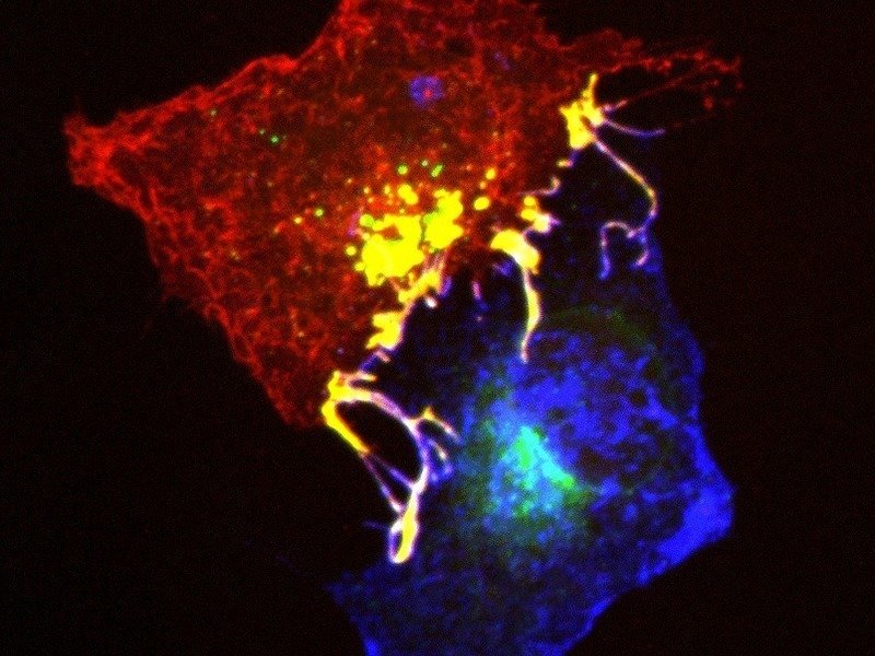
Ephrins (blue) and Ephs (red) form complexes (yellow) at cell contact points.

When cells grow and divide, they come into contact with other cells. This happens not only during development and regeneration and after injury, but also during cancer growth and the formation of metastases. When cells come into contact with each other in this way, information is exchanged by proteins, which are embedded in the cell membranes and form tight lock-and-key complexes with each other. These connections must be severed if the cells want to transmit a repulsion signal. It appears that the fastest way to do this is for the cells to engulf the protein complex from the membrane of the neighbouring cell. Scientists from the Max Planck Institute of Neurobiology in Martinsried have now identified the molecules that control this process.

Development is an extremely rapid process. Increasing numbers of cells are formed which must find their correct position in the body, clearly demarcate themselves from each other to form tissue, or – as is the case in the nervous system – establish contact with partner cells in remote locations. “The crowding is accompanied by orderly pushing and shoving,” says Rüdiger Klein, whose Department at the Max Planck Institute of Neurobiology studies how cells get their bearings. “A popular way for one cell to show another which direction to take is for it to repel the other cell following brief contact.” According to the scientists’ observations, the cells do not exactly treat each other with kid gloves and even go so far as to engulf entire pieces from the membranes of other cells. More


Kank2 expression pattern

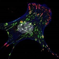
Tissue are composed of cells and their surrounding extracellular matrix. During embryonic development, wound healing and cancer metastasis, cells move within tissues. How they do it, is a highly relevant scientific and medical question. Now, researcher at the Max Planck Institute of Biochemistry in Martinsried found a new molecule that regulates the speed of cell migration. Kank2, the newly discovered protein, diminishes the force transmission between cells and extracellular matrix. Thereby the cells find less grip on the ground and move slower.

In tissue, cells are embedded within the extracellular matrix, a meshwork of different protein assemblies. Integrins are anchor proteins on the cell surface, which on the one hand enable cells to adhere to the extracellular matrix, and on the other hand connect to the actin cytoskeleton inside the cells. Hence, integrin-mediated adhesion points transmit mechanical force generated by the contractile cytoskeleton to the extracellular matrix, which is needed for cell migration. The stronger integrin-based adhesions are pulled by mechanical force, the tighter they are anchored on the extracellular matrix. More


The adult brain has learned to calculate an image of its environment from sensory information. If the input signals change, however, even the adult brain is able to adapt − and, ideally, to return to its original activity patterns once the perturbation has ceased. Scientists at the Max Planck Institute of Neurobiology in Martinsried have now shown in mice that this ability is due to the properties of individual neurons. Their findings demonstrate that individual cells adjust strongly to changes in the environment but after the environment returns to its original state it is again the individual neurons which reassume their initial response properties. This could explain why despite substantial plasticity the perception in the adult brain is rather stable and why the brain does not have to continuously relearn everything.

Everything we know about our environment is based on calculations in our brain. Whereas a child's brain first has to learn the rules that govern the environment, the adult brain knows what to expect and, for the most part, processes environmental stimuli in a stable manner. Yet even the adult brain is able to respond to changes, to form new memories and to learn. Research in recent years has shown that changes to the connections between neurons form the basis of this plasticity. But, how can the brain continually change its connections and learn new things without jeopardizing its stable representation of the environment? Neurobiologists in the Department of Tobias Bonhoeffer in Martinsried have now addressed this fundamental question and looked at the interplay between plasticity and stability.

The scientists studied the stability of the processing of sensations in the visual cortex of the mouse. It has been known for about 50 years that when one eye is temporarily closed, the region of the brain responsive to that eye increasingly becomes responsive to signals from the other eye that is still open. This insight has been important to optimize the use of eye patches in children with a squint. “Thanks to new genetically encoded indicators, it has recently become possible to observe reliably the activity of individual neurons over long periods of time,” says Tobias Rose, the lead author of the study. “With a few additional improvements, we were able to show for the first time what happens in the brain on the single-cell level when such environmental changes occur.”

read more


F.-Ulrich Hartl, director at the Max Planck Institute of Biochemistry, will receive, together with Arthur L. Horwich and Susan L. Lindquist, this year’s Albany Medical Center Prize. The Albany Medical and Research Prize is endowed with $ 500,000 (450,000 €) and is one of America’s most distinguished Prizes in Medicine. The three awardees were honored for their fundamental and complementary discoveries related to the mechanism of protein folding. The prize will be awarded on September 28 in Albany, NY, USA.

Proteins are small molecular machines within each cell that perform a wide variety of essential tasks. To carry out their critical functions, newly produced immature chain-like proteins have to fold into specific, three-dimensional structures. In the 1980s F.-Ulrich Hartl and Arthur L. Horwich proved that proteins do not fold spontaneously, as had previously been thought. Rather, they need assistants in their folding process, so called chaperones. Hartl and Horwich discovered that certain chaperones function as cage-like ‘folding machines’, offering newly synthesized proteins a protected environment, which allows them to fold properly and adopt their appropriate functional structures. more


Computation of motion by T4 cells in the fly brain is more complex than previously believed. As indicated by their name, photoreceptor cells in the eye respond to light: is an image point bright or dark? They do not indicate the direction of a movement. This perception only arises in the brain through the comparative computations of light signals coming from adjacent image points. Engineers, physicists and neurobiologists have been debating the exact nature of these computations for around 50 years.
Scientists from the Max Planck Institute of Neurobiology have now combined two theories about these computations, which were previously considered to be alternative hypotheses – and discovered that they are carried out in a single neuron.Flies are usually very difficult to catch. No wonder – they invest around ten percent of their brain in the detection and processing of image motion. For the fly, a hand approaches in slow motion and the fly’s evasive manoeuvre has long been triggered before any real danger arises. Scientists have been researching for decades how the fly brain can perceive and process movements so quickly and accurately. “Our goal is slowly coming into view, and we are close to completely decoding the neuronal circuit of motion perception in the fly,” says Alexander Borst, who has been working on this problem with his Department at the Max Planck Institute of Neurobiology for quite some time. The scientists have now come one step closer to the answer: They have provided experimental data that combine two theories previously considered as alternatives.  More


Do you speak -omics? If you don't, Perseus ­– www.perseus-framework.org might be able to help you. Researchers from the Max Planck Institute in Martinsried have developed this free software platform for users of high-throughput techniques, such as mass spectrometry, in order to translate raw biological data into relevant findings. As reported in the current issue of Nature Methods, molecular signatures from cells, tissue and body fluids can be identified and characterized on this platform without the need for bioinformatic training. Perseus was designed to deal with proteomic studies in which data on thousands of proteins is processed. It has, however, also proven itself in other molecular studies and will be expanded accordingly.
Absolutely nothing in an organism works without proteins. These molecules operate as molecular machines, act as building materials and appear in a variety of other roles. However, they are rarely lone warriors, with the result that analyzing the sum total of all proteins in a cell, a tissue, a body fluid or even in an entire organism is essential. This can establish when and where a particular molecule appears in what quantity and with whom it interacts. Corresponding approaches exist for other biological molecules as well. Modern high-throughput techniques such as mass spectrometry provide the necessary raw data, often from several thousand different proteins. More


Publication Placeholder

Spadaro, M., Gerdes, L.A., Krumbholz, M., Ertl-Wagner, B., Thaler, F.S., Schuh, E., Metz, I., Blaschek, A., Dick, A., Bruck, W., Hohlfeld, R., Meinl, E., and Kumpfel, T.
Neurol Neuroimmunol Neuroinflamm, 2016, 3, e257.

Autoantibodies to MOG in a distinct subgroup of adult multiple sclerosis.

OBJECTIVES: To evaluate the presence of antibodies to conformation-intact myelin oligodendrocyte glycoprotein (MOG) in a subgroup of adult patients with clinically definite multiple sclerosis (MS) preselected for a specific clinical phenotype including severe spinal cord, optic nerve, and brainstem involvement.

METHODS: Antibodies to MOG were investigated using a cell-based assay in 3 groups of patients: 104 preselected patients with MS (group 1), 55 age- and sex-matched, otherwise unselected patients with MS (group 2), and in 22 brain-biopsied patients with demyelinating diseases of the CNS (n = 19 with MS), 4 of whom classified as MS type II (group 3). Recognized epitopes were identified with mutated variants of MOG.

RESULTS: Antibodies to MOG were found in about 5% (5/104) of preselected adult patients with MS. In contrast, in groups 2 and 3, none of the patients tested positive for MOG antibodies. Patients with MS with antibodies to MOG predominantly manifested with concomitant severe brainstem and spinal cord involvement and had a severe disease course with high relapse rates and failure to several disease-modifying therapies. Three of them had been treated with plasma exchange with a favorable response. All anti-MOG-positive patients with MS showed typical MS lesions on brain MRI. Longitudinal analysis up to 9 years revealed fluctuations and reappearance of anti-MOG reactivity. Epitope mapping indicated interindividual heterogeneity, yet intraindividual stability of the antibody response.

CONCLUSIONS: Antibodies to MOG can be found in a distinct subgroup of adult MS with a specific clinical phenotype and may indicate disease heterogeneity.