News

Publication Placeholder

Lemke, S.B., Weidemann, T., Cost, A.L., Grashoff, C., and Schnorrer, F.
PLoS Biol, 2019, 17, e3000057.
doi: 10.1371/journal.pbio.3000057

A small proportion of Talin molecules transmit forces at developing muscle attachments in vivo.

Cells in developing organisms are subjected to particular mechanical forces that shape tissues and instruct cell fate decisions. How these forces are sensed and transmitted at the molecular level is therefore an important question, one that has mainly been investigated in cultured cells in vitro. Here, we elucidate how mechanical forces are transmitted in an intact organism. We studied Drosophila muscle attachment sites, which experience high mechanical forces during development and require integrin-mediated adhesion for stable attachment to tendons. Therefore, we quantified molecular forces across the essential integrin-binding protein Talin, which links integrin to the actin cytoskeleton. Generating flies expressing 3 Förster resonance energy transfer (FRET)-based Talin tension sensors reporting different force levels between 1 and 11 piconewton (pN) enabled us to quantify physiologically relevant molecular forces. By measuring primary Drosophila muscle cells, we demonstrate that Drosophila Talin experiences mechanical forces in cell culture that are similar to those previously reported for Talin in mammalian cell lines. However, in vivo force measurements at developing flight muscle attachment sites revealed that average forces across Talin are comparatively low and decrease even further while attachments mature and tissue-level tension remains high. Concomitantly, the Talin concentration at attachment sites increases 5-fold as quantified by fluorescence correlation spectroscopy (FCS), suggesting that only a small proportion of Talin molecules are mechanically engaged at any given time. Reducing Talin levels at late stages of muscle development results in muscle-tendon rupture in the adult fly, likely as a result of active muscle contractions. We therefore propose that a large pool of adhesion molecules is required to share high tissue forces. As a result, less than 15% of the molecules experience detectable forces at developing muscle attachment sites at the same time. Our findings define an important new concept of how cells can adapt to changes in tissue mechanics to prevent mechanical failure in vivo.

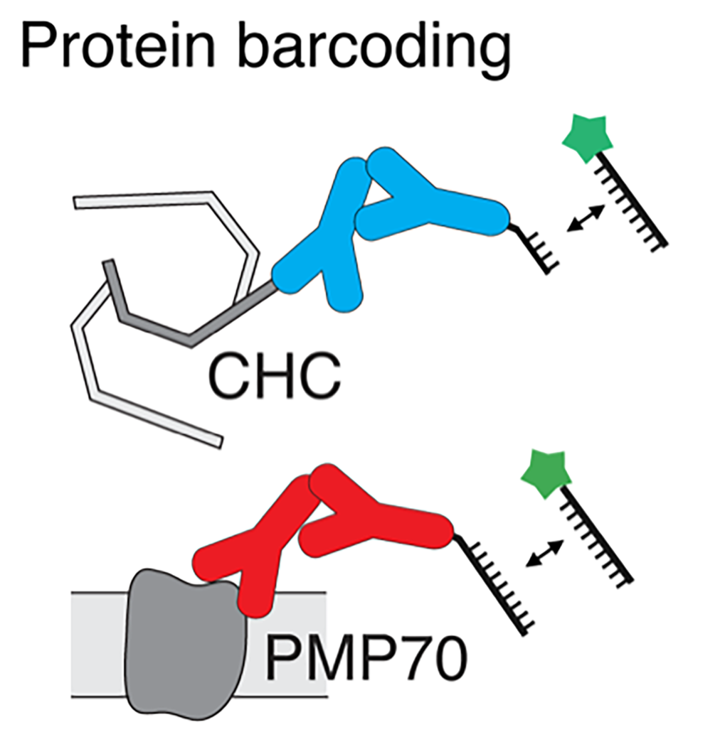
In everyday life, blinking lights can send signals – for example, that a car is going to turn. Now, researchers have engineered tiny “blinkers” that reveal single molecules of RNA or protein inside cells based on the duration and frequency of each flash. The research, published in the ACS journal Nano Letters, could allow scientists to see the locations of many different biomolecules in a cell simultaneously, possibly leading to better diagnostics and treatments.

Recently, scientists have developed super-resolution microscopes that can image single molecules that are only a few nanometers in size. To discriminate a specific nucleic acid or protein, they typically add a fluorescent probe that binds to that molecule and emits a certain wavelength of light. However, because the emission wavelengths of different fluorescent probes can overlap, researchers can usually only detect three or four unique proteins or nucleic acids at a time, instead of the thousands that exist in cells. Ralf Jungmann and colleagues from the Max Planck Institute of Biochemistry in Martinsried wondered if they could use fluorescent probes that blink with light at a variable duration and frequency to detect dozens of biomolecules at once. That way, they could use a single fluorophore to image many different molecules.

The researchers based their system on complementary sequences of DNA that come together to link a fluorophore with a target biomolecule and then fall apart again, generating a blinking fluorescent signal. By varying the length and number of DNA sequences bound to the target, the researchers could adjust how long the blink lasted, as well as how often blinks occurred. To test their approach in cells, the researchers imaged two different RNA molecules and two proteins. Then, they used three fluorescent probes to image 124 distinct DNA structures that contained different numbers of target DNAs so that they blinked at different frequencies. The procedure took only a few minutes and had an accuracy of 97.6 percent, the researchers say.

read more


graduation

Congratulations on your PhD!

Leonie Mönkemeyer
Structural and Functional Studies on the Eukaryotic Chaperonin TRiC and its Cooperating Chaperone Hgh1
RG: F.-Ulrich Hartl

Yan Xiao
GroEL Ring Separation and Exchange in the Chaperonin Reaction
RG: F.-Ulrich Hartl


Publication Placeholder

Monkemeyer, L., Klaips, C.L., Balchin, D., Korner, R., Hartl, F.U., and Bracher, A.
Mol Cell, 2019, [Epub ahead of print].
doi: 10.1016/j.molcel.2019.01.034

Chaperone Function of Hgh1 in the Biogenesis of Eukaryotic Elongation Factor 2

Eukaryotic elongation factor 2 (eEF2) is an abundant and essential component of the translation machinery. The biogenesis of this 93 kDa multi-domain protein is assisted by the chaperonin TRiC/CCT. Here, we show in yeast cells that the highly conserved protein Hgh1 (FAM203 in humans) is a chaperone that cooperates with TRiC in eEF2 folding. In the absence of Hgh1, a substantial fraction of newly synthesized eEF2 is degraded or aggregates. We solved the crystal structure of Hgh1 and analyzed the interaction of wild-type and mutant Hgh1 with eEF2. These experiments revealed that Hgh1 is an armadillo repeat protein that binds to the dynamic central domain III of eEF2 via a bipartite interface. Hgh1 binding recruits TRiC to the C-terminal eEF2 module and prevents unproductive interactions of domain III, allowing efficient folding of the N-terminal GTPase module. eEF2 folding is completed upon dissociation of TRiC and Hgh1.


Publication Placeholder

Cost, A.L., Khalaji, S., and Grashoff, C.
Curr Protoc Cell Biol, 2019, e85, [Epub ahead of print].
doi: 10.1002/cpcb.85

Genetically Encoded FRET-Based Tension Sensors

Genetically encoded Förster resonance energy transfer (FRET)-based tension sensors measure piconewton-scale forces across individual molecules in living cells or whole organisms. These biosensors show comparably high FRET efficiencies in the absence of tension, but FRET quickly decreases when forces are applied. In this article, we describe how such biosensors can be generated for a specific protein of interest, and we discuss controls to confirm that the observed differences in FRET efficiency reflect changes in molecular tension. These FRET efficiency changes can be related to mechanical forces as the FRET-force relationship of the employed tension sensor modules are calibrated. We provide information on construct generation, expression in cells, and image acquisition using live-cell fluorescence lifetime imaging microscopy (FLIM). Moreover, we describe how to analyze, statistically evaluate, and interpret the resulting data sets. Together, these protocols should enable the reader to plan, execute, and interpret FRET-based tension sensor experiments.


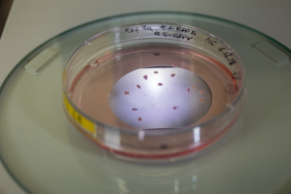
Much insight into the brain’s function has been gained from scientists working with animal models. However, animal models have their limitations since animal brains undergo a very different development process to humans brains. Some research has been carried out on the human brain, but it has relied on post-mortem donations and cultured cells. Since human brain tissue is difficult to obtain and animal models can only model the human brain to a limited extent, there was a crucial gap in brain research and a novel method to model human disease was highly sought after.

The Cappello research group at the Max Planck Institute of Psychiatry in a highly collaborative work, particularly with the laboratory of Barbara Treutlein from the Max Planck Institute for Evolutionary Anthropology in Leipzig, have employed brain organoids to model developmental brain malfunctions. These brain organoids have revolutionized neuroscience as they can recapitulate the way neurons differentiate in development to a remarkably high degree. When the human brain develops, new cells grow and aggregate in a very specific order.

The brain organoids in this study were grown from skin cell biopsies donated by patients. Silvia Cappello, who leads the work, explains: “We reprogrammed the skin cells into induced pluripotent stem cells. These pluripotent stem cells were then used to generate brain cells, which could be differentiated into many different types of brain cells.” The different types of brain cells and their interactions can then be studied behaving in a way they would in an actual human brain. Cappello adds: “Brain organoids give us a much clearer picture of how brain cells are functioning and can accurately model human neurological diseases.”

The ability to model human brain development in vitro holds tremendous translational value. Brain organoids have already helped scientists to better understand the Zika virus, Alzheimer’s disease and autism. As the brain organoid is grown from the cells of an individual, it allows scientists to study exactly what is happening in individual patients. Cappello concludes: “Validating brain organoids represents a hugely important step in helping us to understand developmental and neurological disorders and they hold great promise for discovering new treatments.”

read more


Publication Placeholder

Schopf, F.H., Huber, E.M., Dodt, C., Lopez, A., Biebl, M.M., Rutz, D.A., Muhlhofer, M., Richter, G., Madl, T., Sattler, M., Groll, M., and Buchner, J.
Mol Cell, 2019, [Epub ahead of print].
doi: 10.1016/j.molcel.2019.02.011

The Co-chaperone Cns1 and the Recruiter Protein Hgh1 Link Hsp90 to Translation Elongation via Chaperoning Elongation Factor 2

The Hsp90 chaperone machinery in eukaryotes comprises a number of distinct accessory factors. Cns1 is one of the few essential co-chaperones in yeast, but its structure and function remained unknown. Here, we report the X-ray structure of the Cns1 fold and NMR studies on the partly disordered, essential segment of the protein. We demonstrate that Cns1 is important for maintaining translation elongation, specifically chaperoning the elongation factor eEF2. In this context, Cns1 interacts with the novel co-factor Hgh1 and forms a quaternary complex together with eEF2 and Hsp90. The in vivo folding and solubility of eEF2 depend on the presence of these proteins. Chaperoning of eEF2 by Cns1 is essential for yeast viability and requires a defined subset of the Hsp90 machinery as well as the identified eEF2 recruiting factor Hgh1.


Our world is full of sensory stimuli. Depending on what we see, smell, taste, feel, or hear, we are compelled to behave in a predictable way – like approaching tasty food or avoiding an oncoming car. The brain’s ability to make sense of the diverse sensory stimuli and to coordinate the appropriate behavioral response relies critically on the function of the cerebellum. This hindbrain region, critical to sensorimotor coordination, is conserved across vertebrates, from humans to birds to fish.

The mammalian cerebellum, however, contains hundreds of thousands of Purkinje cells, each receiving inputs from many thousands of presynaptic neurons. Cracking the cerebellar code here is nearly impossible, even with the latest methods. Ruben Portugues and his team thus focus on a "simpler" version: the cerebellum of six to eight day old zebrafish larvae.

“At this age, the zebrafish cerebellum contains about 500 Purkinje cells and is involved in behaviors such as swimming and eye movements”, explains Laura Knogler, who studied the cerebellar circuits together with graduate student Andreas Kist. “It’s all there and still very complex, but we have a chance to see all cells’ activity in the transparent brains of these fish and directly record the detailed activity of individual cells.” By studying the cerebellum of zebrafish larvae behaving within virtual-reality environments, the scientists were now able to tackle the central question of how the cerebellum coordinates behavior.

Like many vertebrates, zebrafish use visual cues to direct their movements, keep track of their environment or to identify potential predators or prey. Using this knowledge, the neuroscientists showed the fish different visual stimuli while observing neuronal activity and the motor functions. The surprising result was a cerebellar division into three behavioral modules, each encoding a distinct type of visual information: directional motion onset, rotational motion velocity, or changes in luminance. Every studied Purkinje cell belonged to one of these three modules.

In contrast, the behavior of the fish was encoded in nearly the same way by all cells. This became visible in an impressive way when the fish were swimming: “Nearly the entire cerebellum lit up with fluorescence, showing an overwhelming amount of Purkinje cell excitation during swim bouts”, relates Andreas Kist. The scientists believe that the observed cerebellar organization is an important trait for neural coding and associative learning: “The modules appeared optimized to organize the information necessary for the principal behaviors of the zebrafish at this age, yet may also allow for the flexibility required to learn new things through experience”, explains Knogler. “I wouldn’t be surprised if other sensory input and the cerebella of other species are organized in a similar way.”.

read more


Publication Placeholder

Bantele, S.C.S., Lisby, M., and Pfander, B.
Nat Commun, 2019, 10, 944.
doi: 10.1038/s41467-019-08889-5

Quantitative sensing and signalling of single-stranded DNA during the DNA damage response

The DNA damage checkpoint senses the presence of DNA lesions and controls the cellular response thereto. A crucial DNA damage signal is single-stranded DNA (ssDNA), which is frequently found at sites of DNA damage and recruits the sensor checkpoint kinase Mec1-Ddc2. However, how this signal - and therefore the cell's DNA damage load - is quantified, is poorly understood. Here, we use genetic manipulation of DNA end resection to induce quantitatively different ssDNA signals at a site-specific double strand break in budding yeast and identify two distinct signalling circuits within the checkpoint. The local checkpoint signalling circuit leading to γH2A phosphorylation is unresponsive to increased amounts of ssDNA, while the global checkpoint signalling circuit, which triggers Rad53 activation, integrates the ssDNA signal quantitatively. The global checkpoint signal critically depends on the 9-1-1 and its downstream acting signalling axis, suggesting that ssDNA quantification depends on at least two sensor complexes.


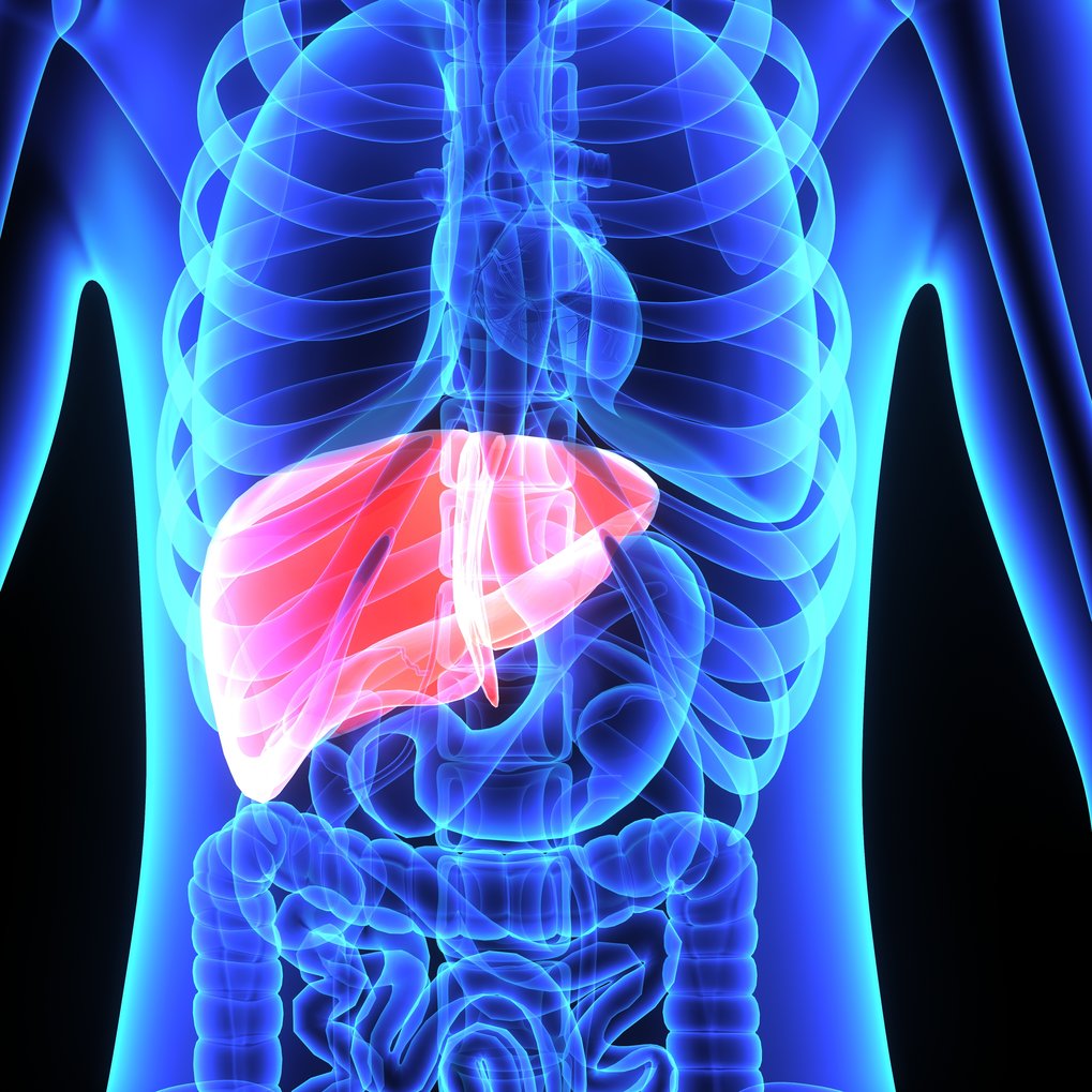
One in four people in Western and Asian societies develop a build-up of fat in the liver as a result of an unhealthy diet. This disease – referred to as non-alcoholic fatty liver disease (NAFLD) – causes no symptoms initially but can develop into end-stage liver cirrhosis with limited treatment options. A discovery, published today in Molecular Systems Biology, paves the way for a simple blood test to detect early stages of NAFLD, opening up the possibility of preventing the development of liver cirrhosis through lifestyle changes or pharmaceutical intervention.

The liver is an important organ, filtering toxic substances from the body and producing proteins required for digestion, blood clotting, and other important physiological functions. “The liver is very resilient and capable of regenerating itself, which may be the reason why liver damages due to excessive fat deposition can go undetected for a long time,” says EMBO Member Matthias Mann of the Max Planck Institute of Biochemistry in Martinsried, Germany, and the University of Copenhagen, Denmark, who led the study. However, when damage accumulates liver function eventually starts to fail.

To date, the standard procedure for diagnosing NAFLD is liver biopsy – a cumbersome and costly procedure that can lead to complications. Non-invasive methods that reliably detect early stage NAFLD are therefore urgently required.

Matthias Mann and his colleagues investigated the plasma proteome – the entire set of proteins present in the blood plasma – of NAFLD patients. Using sophisticated mass spectrometry technologies, they uncovered a set of proteins that accumulate in the plasma of patients with non-symptomatic NAFLD.

read more