Papadopoulou, A.A., Muller, S.A., Mentrup, T., Shmueli, M.D., Niemeyer, J., Haug-Kroper, M., von Blume, J., Mayerhofer, A., Feederle, R., Schroder, B., Lichtenthaler, S.F., and Fluhrer, R.
EMBO Rep, 2019, [Epub ahead of print].
doi: 10.15252/embr.201846451
Signal Peptide Peptidase-Like 2c (SPPL2c) impairs vesicular transport and cleavage of SNARE proteins
Members of the GxGD-type intramembrane aspartyl proteases have emerged as key players not only in fundamental cellular processes such as B-cell development or protein glycosylation, but also in development of pathologies, such as Alzheimer's disease or hepatitis virus infections. However, one member of this protease family, signal peptide peptidase-like 2c (SPPL2c), remains orphan and its capability of proteolysis as well as its physiological function is still enigmatic. Here, we demonstrate that SPPL2c is catalytically active and identify a variety of SPPL2c candidate substrates using proteomics. The majority of the SPPL2c candidate substrates cluster to the biological process of vesicular trafficking. Analysis of selected SNARE proteins reveals proteolytic processing by SPPL2c that impairs vesicular transport and causes retention of cargo proteins in the endoplasmic reticulum. As a consequence, the integrity of subcellular compartments, in particular the Golgi, is disturbed. Together with a strikingly high physiological SPPL2c expression in testis, our data suggest involvement of SPPL2c in acrosome formation during spermatogenesis.
News
Cosolo, A., Jaiswal, J., Csordas, G., Grass, I., Uhlirova, M., and Classen, A.K.
Elife 8, 2019.
doi: 10.7554/eLife.41036
JNK-dependent cell cycle stalling in G2 promotes survival and senescence-like phenotypes in tissue stress
The restoration of homeostasis after tissue damage relies on proper spatial-temporal control of damage-induced apoptosis and compensatory proliferation. In Drosophila imaginal discs these processes are coordinated by the stress response pathway JNK. We demonstrate that JNK signaling induces a dose-dependent extension of G2 in tissue damage and tumors, resulting in either transient stalling or a prolonged but reversible cell cycle arrest. G2-stalling is mediated by downregulation of the G2/M-specific phosphatase String(Stg)/Cdc25. Ectopic expression of stg is sufficient to suppress G2-stalling and reveals roles for stalling in survival, proliferation and paracrine signaling. G2-stalling protects cells from JNK-induced apoptosis, but under chronic conditions, reduces proliferative potential of JNK-signaling cells while promoting non-autonomous proliferation. Thus, transient cell cycle stalling in G2 has key roles in wound healing but becomes detrimental upon chronic JNK overstimulation, with important implications for chronic wound healing pathologies or tumorigenic transformation.
We don’t need to think twice: if an object is approaching on a collision course, we quickly get out of its way. But if something captures our interest, we move directly towards it. Little is known about how the brain classifies visual objects as either attractive or threatening, and how this information is channeled to initiate an appropriate behavior. This gap in our knowledge is now being filled.
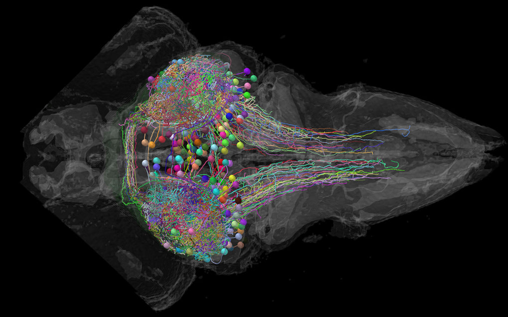
Zebrafish larvae are about five millimeters long and almost transparent, so that we can peek into their brain while it is engaged in a behavioral task. With the aid of newly developed optical and genetic methods, scientists are now able to observe the activity and activation sequence of individual nerve cells. Scientists are thus able to follow the transition from a visual perception to a behavior in real time under the microscope. What the neurobiologists discovered is that “predator” or “prey” categories each activate a dedicated nerve tract to steer behavior.
Previous studies had indicated that this activity originates in the tectum of the fish brain. Humans also have such a tectum, the superior colliculus, which is thought to have very similar functions. To understand what happens in the fish tectum at the cellular level, Thomas Helmbrecht from the Max Planck Institute of Neurobiology studied the reaction among young fish to virtual dots, while observing the activity of their nerve cells and manipulating them using optogenetic methods.
Depending on the size and animated movement, the dots were initially classified as prey or predator in the tectum of the zebrafish. The tectum then transmitted the decision made in each case to the hindbrain via one of two different, spatially separate pathways of nerve cells.
The neurons at the end of the signal chain initiated either an avoidance or approach movement, depending on which of the two pathways carried the information. The scientists were also able to demonstrate that the nerve cells transmitted precise data relating to the position of the potential prey via the approach pathway. The muscles can evidently be controlled by the neurons in such a way that the young fish is able to swim directly towards its prey.
At least 29 different nerve cell types in the tectum project information throughout the brain. “We now want to find out in detail how these individual cell types contribute to behavior,” explains Herwig Baier, in whose laboratory the experiments were conducted. “For the first time, we have the opportunity to fully reconstruct the brain activity that forms the basis of a complex behavioral decision, from the sensory input all the way to the motor output.”
Wang, H.*, Yan, X.*, Aigner, H., Bracher, A., Nguyen, N.D., Hee, W.Y., Long, B.M., Price, G.D., Hartl, F.U., and Hayer-Hartl, M.
Nature, 2019, [Epub ahead of print].
*equal contribution
doi: 10.1038/s41586-019-0880-5
Rubisco condensate formation by CcmM in beta-carboxysome biogenesis
Cells use compartmentalization of enzymes as a strategy to regulate metabolic pathways and increase their efficiency. The α- and β-carboxysomes of cyanobacteria contain ribulose-1,5-bisphosphate carboxylase/oxygenase (Rubisco)-a complex of eight large (RbcL) and eight small (RbcS) subunits-and carbonic anhydrase. As HCO3- can diffuse through the proteinaceous carboxysome shell but CO2 cannot, carbonic anhydrase generates high concentrations of CO2 for carbon fixation by Rubisco. The shell also prevents access to reducing agents, generating an oxidizing environment. The formation of β-carboxysomes involves the aggregation of Rubisco by the protein CcmM, which exists in two forms: full-length CcmM (M58 in Synechococcus elongatus PCC7942), which contains a carbonic anhydrase-like domain followed by three Rubisco small subunit-like (SSUL) modules connected by flexible linkers; and M35, which lacks the carbonic anhydrase-like domain. It has long been speculated that the SSUL modules interact with Rubisco by replacing RbcS. Here we have reconstituted the Rubisco-CcmM complex and solved its structure. Contrary to expectation, the SSUL modules do not replace RbcS, but bind close to the equatorial region of Rubisco between RbcL dimers, linking Rubisco molecules and inducing phase separation into a liquid-like matrix. Disulfide bond formation in SSUL increases the network flexibility and is required for carboxysome function in vivo. Notably, the formation of the liquid-like condensate of Rubisco is mediated by dynamic interactions with the SSUL domains, rather than by low-complexity sequences, which typically mediate liquid-liquid phase separation in eukaryotes. Indeed, within the pyrenoids of eukaryotic algae, the functional homologues of carboxysomes, Rubisco adopts a liquid-like state by interacting with the intrinsically disordered protein EPYC1. Understanding carboxysome biogenesis will be important for efforts to engineer CO2-concentrating mechanisms in plants.
Parhizkar, S., Arzberger, T., Brendel, M., Kleinberger, G., Deussing, M., Focke, C., Nuscher, B., Xiong, M., Ghasemigharagoz, A., Katzmarski, N., Krasemann, S., Lichtenthaler, S.F., Muller, S.A., Colombo, A., Monasor, L.S., Tahirovic, S., Herms, J., Willem, M., Pettkus, N., Butovsky, O., Bartenstein, P., Edbauer, D., Rominger, A., Erturk, A., Grathwohl, S.A., Neher, J.J., Holtzman, D.M., Meyer-Luehmann, M., and Haass, C.
Nat Neurosci, 2019, [Epub ahead of print].
doi: 10.1038/s41593-018-0296-9
Loss of TREM2 function increases amyloid seeding but reduces plaque-associated ApoE
Coding variants in the triggering receptor expressed on myeloid cells 2 (TREM2) are associated with late-onset Alzheimer's disease (AD). We demonstrate that amyloid plaque seeding is increased in the absence of functional Trem2. Increased seeding is accompanied by decreased microglial clustering around newly seeded plaques and reduced plaque-associated apolipoprotein E (ApoE). Reduced ApoE deposition in plaques is also observed in brains of AD patients carrying TREM2 coding variants. Proteomic analyses and microglia depletion experiments revealed microglia as one origin of plaque-associated ApoE. Longitudinal amyloid small animal positron emission tomography demonstrates accelerated amyloidogenesis in Trem2 loss-of-function mutants at early stages, which progressed at a lower rate with aging. These findings suggest that in the absence of functional Trem2, early amyloidogenesis is accelerated due to reduced phagocytic clearance of amyloid seeds despite reduced plaque-associated ApoE.
Glock, P., Ramm, B., Heermann, T., Kretschmer, S., Schweizer, J., Mucksch, J., Alagoz, G., and Schwille, P.
ACS Synth Biol, 2018, [Epub ahead of print].
(IMPRS-LS students are in bold)
doi: 10.1021/acssynbio.8b00415
Stationary patterns in a two-protein reaction-diffusion system.
Patterns formed by reaction-diffusion mechanisms are crucial for the development or sustenance of most organisms in nature. Patterns include dynamic waves, but are more often found as static distributions, such as animal skin patterns. Yet, a simplistic biological model system to reproduce and quantitatively investigate static reaction-diffusion patterns has been missing so far. Here, we demonstrate that the Escherichia coli Min system, known for its oscillatory behavior between the cell poles, is under certain conditions capable of transitioning to quasi-stationary protein distributions on membranes closely resembling Turing patterns. We systematically titrated both proteins, MinD and MinE, and found that removing all purification tags and linkers from the N-terminus of MinE was critical for static patterns to occur. At small bulk heights, dynamic patterns dominate, such as in rod-shaped microcompartments. We see implications of this work for studying pattern formation in general, but also for creating artificial gradients as downstream cues in synthetic biology applications.
Sonal, Ganzinger, K.A., Vogel, S.K., Mucksch, J., Blumhardt, P., and Schwille, P.
J Cell Sci, 2018, 132, [Epub ahead of print].
(IMPRS-LS students are in bold)
doi: 10.1242/jcs.219899
Myosin-II activity generates a dynamic steady state with continuous actin turnover in a minimal actin cortex.
Dynamic reorganization of the actomyosin cytoskeleton allows fast modulation of the cell surface, which is vital for many cellular functions. Myosin-II motors generate the forces required for this remodeling by imparting contractility to actin networks. However, myosin-II activity might also have a more indirect contribution to cytoskeletal dynamics; it has been proposed that myosin activity increases actin turnover in various cellular contexts, presumably by enhancing disassembly. In vitro reconstitution of actomyosin networks has confirmed the role of myosin in actin network disassembly, but the reassembly of actin in these assays was limited by factors such as diffusional constraints and the use of stabilized actin filaments. Here, we present the reconstitution of a minimal dynamic actin cortex, where actin polymerization is catalyzed on the membrane in the presence of myosin-II activity. We demonstrate that myosin activity leads to disassembly and redistribution in this simplified cortex. Consequently, a new dynamic steady state emerges in which the actin network undergoes constant turnover. Our findings suggest a multifaceted role of myosin-II in the dynamics of the eukaryotic actin cortex. This article has an associated First Person interview with the first author of the paper.
Blumhardt, P., Stein, J., Mucksch, J., Stehr, F., Bauer, J., Jungmann, R., and Schwille, P.
Molecules, 2018, 23, [Epub ahead of print].
(IMPRS-LS students are in bold)
doi: 10.3390/molecules23123165
Photo-Induced Depletion of Binding Sites in DNA-PAINT Microscopy
The limited photon budget of fluorescent dyes is the main limitation for localization precision in localization-based super-resolution microscopy. Points accumulation for imaging in nanoscale topography (PAINT)-based techniques use the reversible binding of fluorophores and can sample a single binding site multiple times, thus elegantly circumventing the photon budget limitation. With DNA-based PAINT (DNA-PAINT), resolutions down to a few nanometers have been reached on DNA-origami nanostructures. However, for long acquisition times, we find a photo-induced depletion of binding sites in DNA-PAINT microscopy that ultimately limits the quality of the rendered images. Here we systematically investigate the loss of binding sites in DNA-PAINT imaging and support the observations with measurements of DNA hybridization kinetics via surface-integrated fluorescence correlation spectroscopy (SI-FCS). We do not only show that the depletion of binding sites is clearly photo-induced, but also provide evidence that it is mainly caused by dye-induced generation of reactive oxygen species (ROS). We evaluate two possible strategies to reduce the depletion of binding sites: By addition of oxygen scavenging reagents, and by the positioning of the fluorescent dye at a larger distance from the binding site.
Scacchetti, A., Brueckner, L., Jain, D., Schauer, T., Zhang, X., Schnorrer, F., van Steensel, B., Straub, T., and Becker, P.B.
Life Sci Alliance, 2018, 1, e201800024.
(IMPRS-LS students are in bold)
doi: 10.26508/lsa.201800024
CHRAC/ACF contribute to the repressive ground state of chromatin
Telomeres and the shelterin complex cap and protect the ends of chromosomes. Telomeres are flanked by the subtelomeric sequences that have also been implicated in telomere regulation, although their role is not well defined. Here, we show that, in Schizosaccharomyces pombe, the telomere-associated sequences (TAS) present on most subtelomeres are hyper-recombinogenic, have metastable nucleosomes, and unusual low levels of H3K9 methylation. Ccq1, a subunit of shelterin, protects TAS from nucleosome loss by recruiting the heterochromatic repressor complexes CLRC and SHREC, thereby linking nucleosome stability to gene silencing. Nucleosome instability at TAS is independent of telomeric repeats and can be transmitted to an intrachromosomal locus containing an ectopic TAS fragment, indicating that this is an intrinsic property of the underlying DNA sequence. When telomerase recruitment is compromised in cells lacking Ccq1, DNA sequences present in the TAS promote recombination between chromosomal ends, independent of nucleosome abundance, implying an active function of these sequences in telomere maintenance. We propose that Ccq1 and fragile subtelomeres co-evolved to regulate telomere plasticity by controlling nucleosome occupancy and genome stability.
van Emden, T.S., Forn, M., Forne, I., Sarkadi, Z., Capella, M., Martin Caballero, L., Fischer-Burkart, S., Bronner, C., Simonetta, M., Toczyski, D., Halic, M., Imhof, A., and Braun, S.
EMBO Rep, 2018, [Epub ahead of print].
(IMPRS-LS students are in bold)
doi: 10.15252/embr.201847181
Shelterin and subtelomeric DNA sequences control nucleosome maintenance and genome stability.
Telomeres and the shelterin complex cap and protect the ends of chromosomes. Telomeres are flanked by the subtelomeric sequences that have also been implicated in telomere regulation, although their role is not well defined. Here, we show that, in Schizosaccharomyces pombe, the telomere-associated sequences (TAS) present on most subtelomeres are hyper-recombinogenic, have metastable nucleosomes, and unusual low levels of H3K9 methylation. Ccq1, a subunit of shelterin, protects TAS from nucleosome loss by recruiting the heterochromatic repressor complexes CLRC and SHREC, thereby linking nucleosome stability to gene silencing. Nucleosome instability at TAS is independent of telomeric repeats and can be transmitted to an intrachromosomal locus containing an ectopic TAS fragment, indicating that this is an intrinsic property of the underlying DNA sequence. When telomerase recruitment is compromised in cells lacking Ccq1, DNA sequences present in the TAS promote recombination between chromosomal ends, independent of nucleosome abundance, implying an active function of these sequences in telomere maintenance. We propose that Ccq1 and fragile subtelomeres co-evolved to regulate telomere plasticity by controlling nucleosome occupancy and genome stability.