News

Publication Placeholder

Modic, M., Grosch, M., Rot, G., Schirge, S., Lepko, T., Yamazaki, T., Lee, F.C.Y., Rusha, E., Shaposhnikov, D., Palo, M., Merl-Pham, J., Cacchiarelli, D., Rogelj, B., Hauck, S.M., von Mering, C., Meissner, A., Lickert, H., Hirose, T., Ule, J., and Drukker, M.
Mol Cell, 2019, [Epub ahead of print].
doi: 10.1016/j.molcel.2019.03.041

Cross-Regulation between TDP-43 and Paraspeckles Promotes Pluripotency-Differentiation Transition

RNA-binding proteins (RBPs) and long non-coding RNAs (lncRNAs) are key regulators of gene expression, but their joint functions in coordinating cell fate decisions are poorly understood. Here we show that the expression and activity of the RBP TDP-43 and the long isoform of the lncRNA Neat1, the scaffold of the nuclear compartment "paraspeckles," are reciprocal in pluripotent and differentiated cells because of their cross-regulation. In pluripotent cells, TDP-43 represses the formation of paraspeckles by enhancing the polyadenylated short isoform of Neat1. TDP-43 also promotes pluripotency by regulating alternative polyadenylation of transcripts encoding pluripotency factors, including Sox2, which partially protects its 3' UTR from miR-21-mediated degradation. Conversely, paraspeckles sequester TDP-43 and other RBPs from mRNAs and promote exit from pluripotency and embryonic patterning in the mouse. We demonstrate that cross-regulation between TDP-43 and Neat1 is essential for their efficient regulation of a broad network of genes and, therefore, of pluripotency and differentiation.


Publication Placeholder

Harpprecht, L., Baldi, S., Schauer, T., Schmidt, A., Bange, T., Robles, M.S., Kremmer, E., Imhof, A., and Becker, P.B.
Nucleic Acids Res, 2019, [Epub ahead of print].
doi: 10.1093/nar/gkz473

A Drosophila cell-free system that senses DNA breaks and triggers phosphorylation signalling.

Preblastoderm Drosophila embryo development is characterized by fast cycles of nuclear divisions. Extracts from these embryos can be used to reconstitute complex chromatin with high efficiency. We now discovered that this chromatin assembly system contains activities that recognize unprotected DNA ends and signal DNA damage through phosphorylation. DNA ends are initially bound by Ku and MRN complexes. Within minutes, the phosphorylation of H2A.V (homologous to γH2A.X) initiates from DNA breaks and spreads over tens of thousands DNA base pairs. The γH2A.V phosphorylation remains tightly associated with the damaged DNA and does not spread to undamaged DNA in the same reaction. This first observation of long-range γH2A.X spreading along damaged chromatin in an in vitro system provides a unique opportunity for mechanistic dissection. Upon further incubation, DNA ends are rendered single-stranded and bound by the RPA complex. Phosphoproteome analyses reveal damage-dependent phosphorylation of numerous DNA-end-associated proteins including Ku70, RPA2, CHRAC16, the exonuclease Rrp1 and the telomer capping complex. Phosphorylation of spindle assembly checkpoint components and of microtubule-associated proteins required for centrosome integrity suggests this cell-free system recapitulates processes involved in the regulated elimination of fatally damaged syncytial nuclei.


Messenger RNAs (mRNAs) are the functional link between the genetic information in the cell nucleus and ribosomes, where proteins are synthesized. The structure of mRNAs can be differentiated into translated and untranslated regions. The translated regions serve as templates for the synthesis of proteins, while the untranslated regions have regulatory functions. The untranslated regions of mRNAs of all higher developed cells – from yeast to plants and humans – contain similar characteristic elements. One such element is the poly(A)-tail: a long chain of adenine molecules, one of the RNA building blocks. These tails are added to the end of the mRNA after their synthesis and fulfil many functions e.g. control stability, translation into proteins and localization of the mRNA. Nascent mRNAs have a long tail of up to several hundred adenines, which is then reduced to a species-specific length by enzymes called deadenylases.

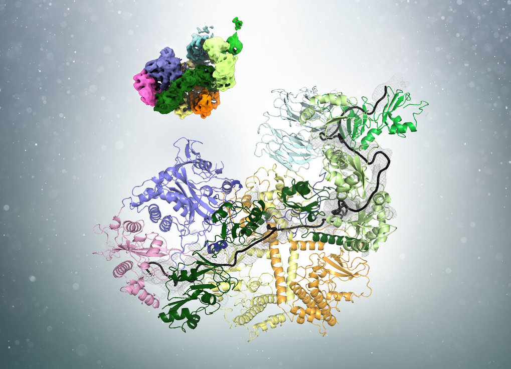
In a recent publication in the journal Cell, researchers led by Elena Conti at the Max Planck Institute of Biochemistry (MPIB) in Martinsried have demonstrated how poly(A)-tail shortening is controlled. “The poly(A)-tail is synthesized to a length much longer than we eventually find in cells. The presence of a poly(A)-tail-trimming mechanism has therefore been long suggested but the details were unclear”, says Ingmar Schäfer, a postdoctoral researcher in Elena Conti’s Department and first author of the study. The researchers now solved the structure of the involved components and show how measuring the length and trimming of the poly(A)-tail are coupled. The process requires the interplay of three components: the poly(A)-tail, poly(A)-tail-binding proteins (PABP) and the Pan2-Pan3 complex of deadenylases.

Using cryo-electron microscopy (cryo-EM), the researchers found that PABP forms arches that cover approximately 25 to 30 bases of the poly(A)-tail. Ingmar Schäfer uses an analogy from everyday life to illustrate the process: “Before cutting hair, a hairdresser uses his fingers to physically determine how much of the hair will be left standing.” Similarly, the PABP arches act as a rulers to determine the length of the poly(A)-tail. “But unlike our hair at the hairdresser, the poly(A)-tail is not trimmed with one cut but rather ‘nibbled off’ from the end by the deadenylase enzyme.”

Interestingly, the number of bound PABP alters the affinity of the deadenylase for the poly(A)-tail. In yeast, the most common poly(A)-tail length is around 30 nucleotides. On longer poly(A)-tails, several PABP can be bound and the adenine chain is quickly degraded. Shorter poly(A)-tails approaching the optimal length can only bind one ruler protein. This corresponds with a lower affinity for the deadenylases. “Hence, PABP is not only the tool measuring the poly(A)-tail, but also acts as a brake on the deadenylases when the optimal length is reached”, explains Schäfer. Eventually, when the mRNA is no longer needed, it is further degraded by a different set of deadenylases.

Polyadenylation is a basic principle of cell biology used to control mRNA stability and protein synthesis. “The structure of the poly(A)-tail ‘trimming tool’ will impact research on a broad variety of aspects of cell biology”, puts MPIB Director Elena Conti the study in a larger context. She highlights that the mechanism is highly conserved between yeasts and humans. “Now that we have solved the structure of the deadenylation machinery in yeast, we want to understand how the system works in human cells, where the optimal poly(A)-tail length differs from yeast.”

read more


Publication Placeholder

van Emden, T.S., and Braun, S.
Curr Genet, 2019, [Epub ahead of print].
doi: 10.1007/s00294-019-00986-8

TASks for subtelomeres: when nucleosome loss and genome instability are favored

Chromosome ends are protected from erosion and chromosomal fusions through telomeric repeats and the telomere-binding protein complex shelterin. Imperfect repetitive sequences, known as telomere-associated sequences (TAS), flank the telomeres, yet their function is not well understood. In this perspective, we discuss our recent findings demonstrating that the TAS, in Schizosaccharomyces pombe, are organized into a distinct chromatin domain that is marked by low nucleosome levels and is highly recombinogenic (van Emden et al. in EMBO Rep 20:e47181). Low nucleosome abundance at the TAS is independent of the chromosomal position, but is an intrinsic property of the DNA sequence itself. Critical nucleosome levels are maintained through two heterochromatin complexes recruited by the shelterin subunit Ccq1, which together control gene repression and nucleosome stability. Furthermore, Ccq1 inhibits TAS-facilitated recombination between subtelomeres, yet independently of nucleosome stability. In conclusion, the TAS present a unique chromatin environment causing nucleosome loss and genome instability, which are both counteracted by Ccq1 through independent mechanisms. Given the antagonistic behavior, we hypothesize that Ccq1 co-evolved with the appearance of TAS to regulate nucleosome dynamics and recombination-based telomere maintenance in the absence of telomerase.


High-grade serous carcinomas (HGSC) make up the majority of ovarian cancer cases. Unfortunately, they have the lowest survival rates. HGSC is a tumor type that occurs primarily in the ovaries and spreads throughout the abdominal cavity. Most patients are diagnosed with late-stage disease that has already spread. Until recently, therapy has been limited to surgery and traditional chemotherapeutic agents. A systematic examination of the tumor and surrounding tissue — particularly normal cells called fibroblasts — has revealed a new treatment target that could potentially prevent the rapid dissemination and poor prognosis associated with high-grade serous carcinoma.

In close collaboration with Fabian Coscia and Matthias Mann, from the Max Planck Institute of Biochemistry in Munich and University of Copenhagen, researchers profiled the expression of more than 5,000 proteins in both normal and cancerous tissues derived from minute amounts of patient biobank material. “When we then got our first data, we were fascinated to find that the metastatic stroma was characterized by a highly conserved protein signature, as opposed to the cancer cells”, adds Fabian Coscia, postdoctoral researcher in Matthias Mann’s group and one of the two first-authors of the study. As these metastatic changes were seen in all of their analyzed patients, the team then went on to understand its functional role during metastasis with the ultimate goal to find novel therapeutic targets.

Indeed, they discovered a metabolic enzyme, nicotinamide N-methyltransferase (NNMT), highly expressed in the stroma surrounding metastatic cancer cells. The researchers found that NNMT causes widespread gene expression changes in the tumor stroma, converting normal fibroblasts to cancer-associated fibroblasts that support and accelerate tumor growth. Stromal NNMT expression encouraged ovarian cancer migration, proliferation, growth and metastasis. It was associated with poor clinical outcomes in patients.

They also found that inhibition of NNMT activity may be able to reduce or even reverse many of the tumor-promoting effects of cancer-associated fibroblasts. This suggests, they note, that the stroma should be explored as a new treatment target. Coscia, co-first author on the manuscript who led the proteomics analysis, added that “this method may be used to discover other proteins that are important for metastasis and to identify early changes during disease development.”

“When we put it all together,” Lengyel added, “it gave us exciting results. We have linked high-end technology including proteomics and metabolomics to functional analysis to improve our understanding of the stroma."

read more


In general, a nerve cell receives input from a number of presynaptic cells, processes the signals, and passes its output to downstream cells. In the cell CT1, however, each of the approximately 1400 cell areas works like a separate neuron. This allows CT1 to access information from all facets of the fly's complex eye and to contribute locally to the calculation of motion direction. Using a computer model of the cell, Alexander Borst and Matthias Meier from the Max Planck Institute of Neurobiology show that CT1 is reaching biophysical limits.

"That’s an amazing cell!" This was the first impression of Alexander Borst, as Matthias Meier showed him the results. Together, the two neurobiologists have demonstrated what is also suspected for amacrine cells in the mammalian retina: It is possible that numerous isolated microcircuits exist in a single nerve cell.

Borst and Meier investigate the visual system of fruit flies, whose complex eyes each consist of about 700 facets. CT1 contacts each of the cell columns that connect to these facets in the brain. In addition, the synapses of CT1 reach into two different brain regions, responsible for the processing of light or dark edges. Thus, CT1 connects to about 1400 areas in the fly brain. This, however, should corrupt the whole system. Each cell column processes changes in light perceived by “their” facet. If the signals of the columns were mixed, the entire image information for downstream cells would be lost.

As flies see very well, a loss of image information does not seem to be an issue. The two neurobiologists could show that each contact area of CT1 is an electrically isolated, independent functional unit. Each of these units receives input from its associated column and returns its output to the same column. Calcium measurements and computer modelling show that essentially, there is no cross-talk between neighboring units or with the cell body.

For the cell units to be electrically isolated from each other, their connections should be thin and long, which increases the electrical resistance. CT1 achieves this with connections of merely 100 nanometers in diameter. In addition, the "connection cables" often form loops. In this way, the connections between neighboring units are about ten times longer than needed to bridge the distance. "It wouldn’t be possible for the connections to get much thinner or longer in the fly brain," says Borst. Why CT1 is so different from most other cells is still a mystery. "It saves cell bodies, but that is certainly not the only reason”, muses Matthias Meier. “If that was the case, such huge amacrine cells wouldn’t be so rare." So far, only very few cells are known with such a structure. Amongst them, CT1 is an extreme example, of which only two cells exist in a fly brain, one per hemisphere.

The scientists are also not yet sure about the exact functions of CT1. The output of the CT1 subunits goes to T4 or T5 cells, depending on their location. These calculate the direction of images moving in front of the fly’s eye. Interestingly, CT1 cells specifically target the motion-sensitive T4 and T5 cells only on one-half of their dendrites. How CT1 thereby affects motion vision is one of the next questions the Max Planck neurobiologists want to investigate.

read more



Publication Placeholder

Hopfler, M., Kern, M.J., Straub, T., Prytuliak, R., Habermann, B.H., Pfander, B., and Jentsch, S.
EMBO J, 2019, [Epub ahead of print].
(IMPRS-LS students are in bold)
doi: 10.15252/embj.2018100368

Slx5/Slx8-dependent ubiquitin hotspots on chromatin contribute to stress tolerance

Chromatin is a highly regulated environment, and protein association with chromatin is often controlled by post-translational modifications and the corresponding enzymatic machinery. Specifically, SUMO-targeted ubiquitin ligases (STUbLs) have emerged as key players in nuclear quality control, genome maintenance, and transcription. However, how STUbLs select specific substrates among myriads of SUMOylated proteins on chromatin remains unclear. Here, we reveal a remarkable co-localization of the budding yeast STUbL Slx5/Slx8 and ubiquitin at seven genomic loci that we term "ubiquitin hotspots". Ubiquitylation at these sites depends on Slx5/Slx8 and protein turnover on the Cdc48 segregase. We identify the transcription factor-like Ymr111c/Euc1 to associate with these sites and to be a critical determinant of ubiquitylation. Euc1 specifically targets Slx5/Slx8 to ubiquitin hotspots via bipartite binding of Slx5 that involves the Slx5 SUMO-interacting motifs and an additional, novel substrate recognition domain. Interestingly, the Euc1-ubiquitin hotspot pathway acts redundantly with chromatin modifiers of the H2A.Z and Rpd3L pathways in specific stress responses. Thus, our data suggest that STUbL-dependent ubiquitin hotspots shape chromatin during stress adaptation.


Publication Placeholder

Burgold, J.*, Schulz-Trieglaff, E.K.*, Voelkl, K., Gutierrez-Angel, S., Bader, J.M., Hosp, F., Mann, M., Arzberger, T., Klein, R., Liebscher, S., and Dudanova, I.
Sci Rep, 2019, 9, 6634.
* authors contributed equally
(IMPRS-LS students and IMPRS-LS alumni are in bold)
doi: 10.1038/s41598-019-43024-w

Cortical circuit alterations precede motor impairments in Huntington's disease mice

Huntington's disease (HD) is a devastating hereditary movement disorder, characterized by degeneration of neurons in the striatum and cortex. Studies in human patients and mouse HD models suggest that disturbances of neuronal function in the neocortex play an important role in disease onset and progression. However, the precise nature and time course of cortical alterations in HD have remained elusive. Here, we use chronic in vivo two-photon calcium imaging to longitudinally monitor the activity of identified single neurons in layer 2/3 of the primary motor cortex in awake, behaving R6/2 transgenic HD mice and wildtype littermates. R6/2 mice show age-dependent changes in cortical network function, with an increase in activity that affects a large fraction of cells and occurs rather abruptly within one week, preceeding the onset of motor defects. Furthermore, quantitative proteomics demonstrate a pronounced downregulation of synaptic proteins in the cortex, and histological analyses in R6/2 mice and human HD autopsy cases reveal a reduction in perisomatic inhibitory synaptic contacts on layer 2/3 pyramidal cells. Taken together, our study provides a time-resolved description of cortical network dysfunction in behaving HD mice and points to disturbed excitation/inhibition balance as an important pathomechanism in HD.


A collaboration between Georgia Tech and the Max Planck Institute of Neurobiology (MPIN) has received a grant of $750,000 over three years from the Human Frontier Science Program (HFSP). The award will allow research on the molecular and genetic encoding of complex behaviors.

The team is led by Georgia Tech’s J. Todd Streelman and MPIN’s Herwig Baier. Streelman is a professor in, and the chair of, the Georgia Tech School of Biological Sciences. Baier is professor and director at the Max Planck Institute of Neurobiology. “It remains incredibly difficult to identify the cellular basis and the genetic variants underlying complex behavior,” Streelman says. “Understanding how behavior is encoded requires solving a dual problem involving neurodevelopment and circuit function.”

To find answers, Streelman and Baier will develop a model system to chart the complex path from genome to brain to behavior in cichlid fish from Lake Malawi. Male cichlid fish build bowers to attract females for mating. The bowers are either pits, which are depressions in the sand, or castles, which look like volcanoes. Each type corresponds to a specific behavior encoded in a fish strain.

When the two strains mate, their male offspring display a remarkable behavior: First they construct a pit then a castle. This behavior indicates that a single brain containing two genomes can produce each behavior in succession. Moreover, gene expression in the brain is biased toward the pit variant of the genome – or pit allele -- when the fish are digging pits and toward the castle allele when they are building castles. “This phenomenon offers the chance to identify both the genome regulatory logic and the neural circuitry underlying complex behavior in one sweep,” Baier says.

Streelman’s group will use single-cell RNA sequencing to pinpoint the cell populations that mediate context-dependent, allele-specific expression in male bower builders. Baier’s team will use genome editing and optogenetic tools to manipulate particular neurons in the brains of behaving bower builders.

The award is one of only 25 made from a total of 654 letters of intent HFSP received from around the world. HFSP provides funding for frontier research in the life sciences. The highly competitive program is implemented by the International Human Frontier Science Program Organization, based in Strasbourg, France.

read more


graduation

Congratulations on your PhD!

Sara Gutiérrez Ángel
Mutant Huntingtin toxicity modifiers revealed by a spatiotemporal proteomic profiling
RG: Rüdiger Klein

Ryan Sherrard
Post-transcriptional regulation of the central apoptotic pathway by microRNAs and RNA-binding proteins during C. elegans development
RG: Barbara Conradt