News

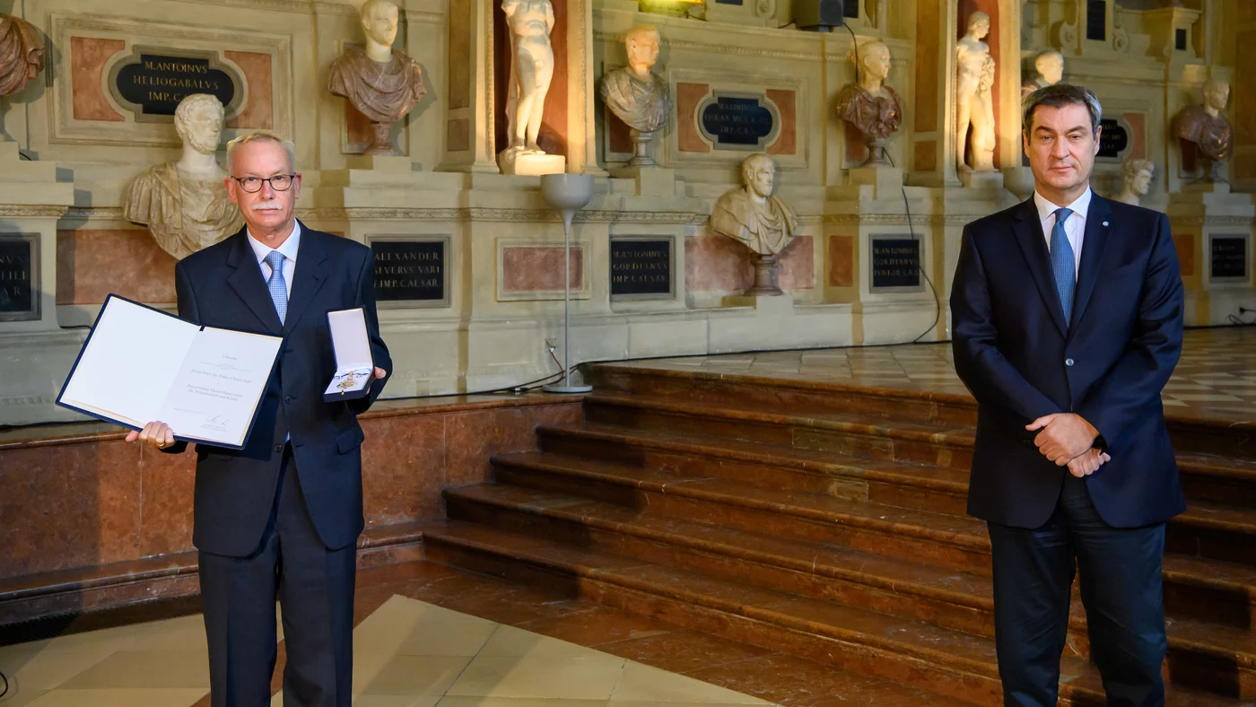
F. Ulrich Hartl, Director at the Max Planck Institute of Biochemistry, together with his colleague Arthur L. Horwich, receives the HFSP Nakasone Prize 2022.

Biochemist F. Ulrich Hartl, together with his colleague and geneticist Arthur L. Horwich, discovered that nascent proteins often do not fold spontaneously into their functional form. Proteins which assist the folding process, known as chaperones, are needed for this process. Both researchers now receive the HFSP Nakasone Prize 2022 of the Human Frontier Science Program for this fundamental and far-reaching discovery. F. Ulrich Hartl: " I am very honored to receive this award together with my early collaborator Art Horwich and look forward to the prize ceremony in Paris." The award honors scientists for their groundbreaking discoveries in areas of the life sciences.

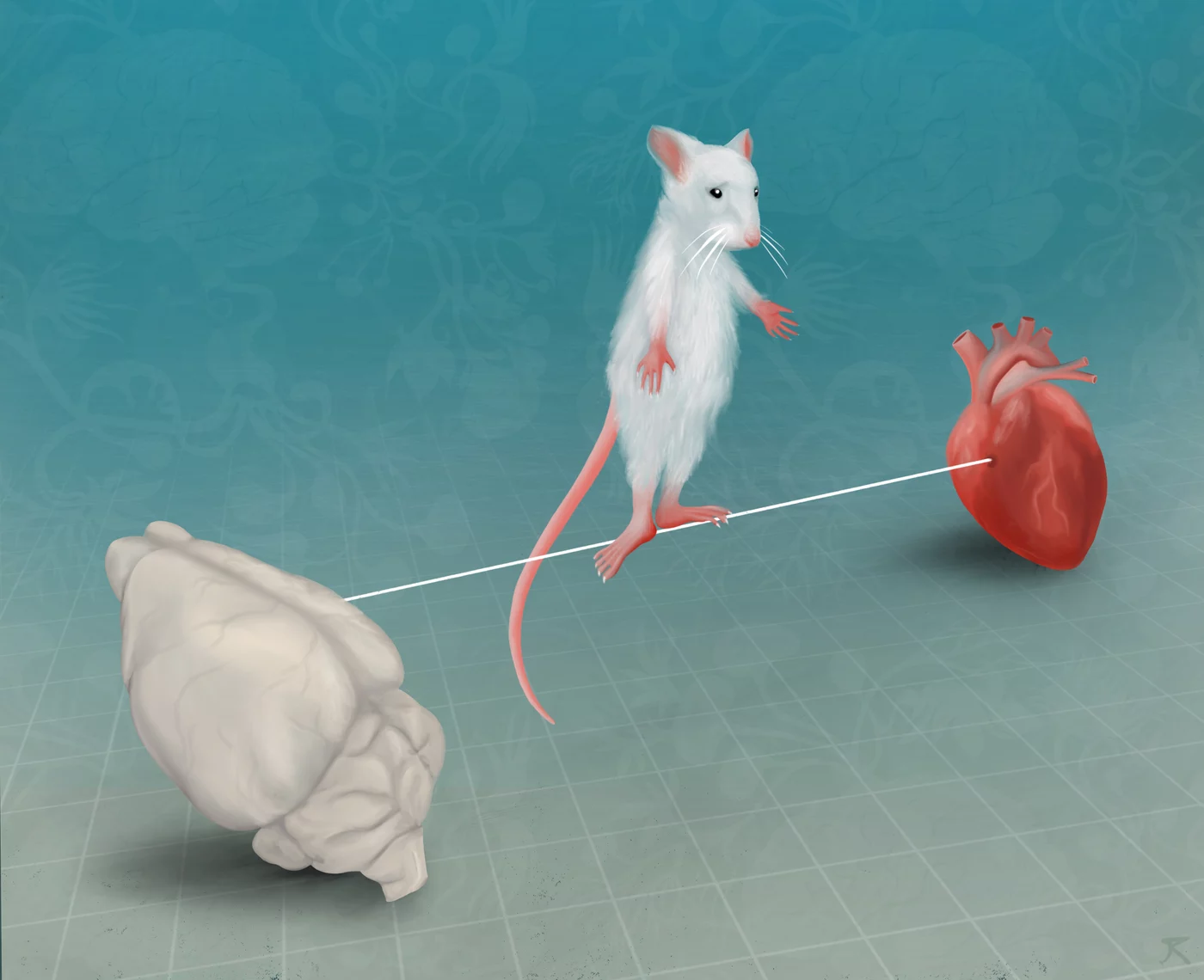
Many proteins need help to fold into their functional form. Helper proteins, known as chaperones, perform this task. This process of assisted folding was discovered by Prof. F. Ulrich Hartl, Director at the Max Planck Institute of Biochemistry in Martinsried, together with his colleague, the American Arthur L. Horwich from the Howard Hughes Medical Institute in Yale, USA. Hartl and his team have been investigating the structure and function of molecular chaperones ever since. Protein aggregations, which are associated with many neurodegenerative diseases such as Parkinson's, Alzheimer's, and Huntington's chorea, can be traced back to malfunctions of the chaperones, among other things. The detailed knowledge of molecular functions and malfunctions of the folding helpers should enable the development of new therapeutic approaches. 

Read more


Publication Placeholder

Markov, D.A., Petrucco, L., Kist, A.M., and Portugues, R.
Nat Commun, 2021, 12, 6694.
(IMPRS-LS students are in bold)
doi: 10.1038/s41467-021-26988-0

A cerebellar internal model calibrates a feedback controller involved in sensorimotor control

Animals must adapt their behavior to survive in a changing environment. Behavioral adaptations can be evoked by two mechanisms: feedback control and internal-model-based control. Feedback controllers can maintain the sensory state of the animal at a desired level under different environmental conditions. In contrast, internal models learn the relationship between the motor output and its sensory consequences and can be used to recalibrate behaviors. Here, we present multiple unpredictable perturbations in visual feedback to larval zebrafish performing the optomotor response and show that they react to these perturbations through a feedback control mechanism. In contrast, if a perturbation is long-lasting, fish adapt their behavior by updating a cerebellum-dependent internal model. We use modelling and functional imaging to show that the neuronal requirements for these mechanisms are met in the larval zebrafish brain. Our results illustrate the role of the cerebellum in encoding internal models and how these can calibrate neuronal circuits involved in reactive behaviors depending on the interactions between animal and environment.


Publication Placeholder

Klein, A.S., Dolensek, N., Weiand, C., and Gogolla, N.
Science, 2021, 374, 1010-1015.
 doi: 10.1126/science.abj8817

Fear balance is maintained by bodily feedback to the insular cortex in mice

How does the brain maintain fear within an adaptive range? We found that the insular cortex acts as a state-dependent regulator of fear that is necessary to establish an equilibrium between the extinction and maintenance of fear memories in mice. Whereas insular cortex responsiveness to fear-evoking cues increased with their certainty to predict harm, this activity was attenuated through negative bodily feedback that arose from heart rate decelerations during freezing. Perturbation of body-brain communication by vagus nerve stimulation disrupted the balance between fear extinction and maintenance similar to insular cortex inhibition. Our data reveal that the insular cortex integrates predictive sensory and interoceptive signals to provide graded and bidirectional teaching signals that gate fear extinction and illustrate how bodily feedback signals are used to maintain fear within a functional equilibrium.


Fear is essential for survival, but must be well regulated to avoid harmful behaviors such as panic attacks or exaggerated risk taking. Scientists at the Max Planck Institute of Neurobiology have now demonstrated in mice that the brain relies on the body’s feedback to regulate fear. The brain’s insular cortex strongly reacts to stimuli signaling danger. However, when the body freezes in response to fear, the heartbeat slows down leading to attenuated insular cortex activity. Processing these opposing signals helps the insular cortex to keep fear in balance. The body’s reactions are thus actively used to regulate emotions and are much more than passive emotional responses.

We usually experience fear as extremely unpleasant. Nevertheless, this emotion has a crucial function: it prevents us from engaging in too risky behaviors. However, this only works if fear is kept within a healthy range. Too intense fear can severely impair our daily lives, as in the case of anxiety disorders or panic attacks. So how can fear be kept in balance? It seems obvious that bodily signals may play a crucial role, as fear causes noticeable changes in our bodies: the heart beats faster or breathing becomes shallower. However, how exactly the brain processes this information to ultimately regulate emotions like fear is still largely unknown. 

Read more


Publication Placeholder

Chrustowicz, J., Sherpa, D., Teyra, J., Siong Loke, M., Popowicz, G., Basquin, J., Sattler, M., Rajan Prabu, J., Sidhu, S.S., and Schulman, B.A.
J Mol Biol,2021, 167347.
doi: 10.1016/j.jmb.2021.167347

Multifaceted N-degron recognition and ubiquitylation by GID/CTLH E3 ligases

N-degron E3 ubiquitin ligases recognize specific residues at the N-termini of substrates. Although molecular details of N-degron recognition are known for several E3 ligases, the range of N-terminal motifs that can bind a given E3 substrate binding domain remains unclear. Here, we discovered capacity of Gid4 and Gid10 substrate receptor subunits of yeast "GID"/human "CTLH" multiprotein E3 ligases to tightly bind a wide range of N-terminal residues whose recognition is determined in part by the downstream sequence context. Screening of phage displaying peptide libraries with exposed N-termini identified novel consensus motifs with non-Pro N-terminal residues binding Gid4 or Gid10 with high affinity. Structural data reveal that conformations of flexible loops in Gid4 and Gid10 complement sequences and folds of interacting peptides. Together with analysis of endogenous substrate degrons, the data show that degron identity, substrate domains harboring targeted lysines, and varying E3 ligase higher-order assemblies combinatorially determine efficiency of ubiquitylation and degradation.


F. Ulrich Hartl, Director at the Max Planck Institute of Biochemistry in Martinsried was awarded the Bavarian Maximilian Order for Science and Art.

F. Ulrich Hartl, director of the research department "Cellular Biochemistry" at the Max Planck Institute of Biochemistry in Martinsried, received the Bavarian Order of Maximilian in the Munich Residence on November 10. With this award, the Free State of Bavaria honors the highest scientific and artistic achievements. F. Ulrich Hartl investigates how proteins fold properly and what role misfolded structures and protein aggregates play in aging and neurodegenerative diseases such as Alzheimer's and Parkinson's: "I feel very honored to receive this recognition." In addition to Hartl, nine other personalities received the award this year, including the president of the Max Planck Society, Martin Stratmann.

Proteins can only fulfill their complex tasks in the organism if they are folded correctly. However, correct folding often requires helper proteins, the so-called molecular chaperones. The researchers led by F. Ulrich Hartl are investigating the structure and function of these molecules. For the "chaperonin" subgroup, the scientists have already been able to show in detail how they work: They encapsulate proteins during their folding so that aggregates cannot form. Such aggregates can be the cause of neurodegenerative diseases such as Alzheimer's, Parkinson's and Huntington's disease. F. Ulrich Hartl and his team are also searching for strategies to prevent proteins from forming aggregates, which one day might become useful in treating these presently incurable diseases. 

Read more


Publication Placeholder

Holtkamp, S.J., Ince, L.M., Barnoud, C., Schmitt, M.T., Sinturel, F., Pilorz, V., Pick, R., Jemelin, S., Mühlstädt, M., Boehncke, W.H., Weber, J., Laubender, D., Philippou-Massier, J., Chen, C.S., Holtermann, L., Vestweber, D., Sperandio, M., Schraml, B.U., Halin, C., Dibner, C., Oster, H., Renkawitz, J., and Scheiermann, C.
(IMPRS-LS students are in bold)
Nat Immunol, 2021, 22, 1375-1381.
doi: 10.1038/s41590-021-01040-x

Circadian clocks guide dendritic cells into skin lymphatics

Migration of leukocytes from the skin to lymph nodes (LNs) via afferent lymphatic vessels (LVs) is pivotal for adaptive immune responses1,2. Circadian rhythms have emerged as important regulators of leukocyte trafficking to LNs via the blood3,4. Here, we demonstrate that dendritic cells (DCs) have a circadian migration pattern into LVs, which peaks during the rest phase in mice. This migration pattern is determined by rhythmic gradients in the expression of the chemokine CCL21 and of adhesion molecules in both mice and humans. Chronopharmacological targeting of the involved factors abrogates circadian migration of DCs. We identify cell-intrinsic circadian oscillations in skin lymphatic endothelial cells (LECs) and DCs that cogovern these rhythms, as their genetic disruption in either cell type ablates circadian trafficking. These observations indicate that circadian clocks control the infiltration of DCs into skin lymphatics, a process that is essential for many adaptive immune responses and relevant for vaccination and immunotherapies.


graduation

Congratulations on your PhD!


Johanna Brüggenthies


Genetic and chemical perturbation of amino acid sening by the GCN1-GCN2 pathway


RG: Peter Murray


Publication Placeholder

Kostrhon, S., Prabu, J.R., Baek, K., Horn-Ghetko, D., von Gronau, S., Klügel, M., Basquin, J., Alpi, A.F., and Schulman, B.A.
(IMPRS-LS students are in bold)
Nat Chem Biol, 2021, 17, 1075-1083.
doi: 10.1038/s41589-021-00858-8

CUL5-ARIH2 E3-E3 ubiquitin ligase structure reveals cullin-specific NEDD8 activation

An emerging mechanism of ubiquitylation involves partnering of two distinct E3 ligases. In the best-characterized E3-E3 pathways, ARIH-family RING-between-RING (RBR) E3s ligate ubiquitin to substrates of neddylated cullin-RING E3s. The E3 ARIH2 has been implicated in ubiquitylation of substrates of neddylated CUL5-RBX2-based E3s, including APOBEC3-family substrates of the host E3 hijacked by HIV-1 virion infectivity factor (Vif). However, the structural mechanisms remained elusive. Here structural and biochemical analyses reveal distinctive ARIH2 autoinhibition, and activation on assembly with neddylated CUL5-RBX2. Comparison to structures of E3-E3 assemblies comprising ARIH1 and neddylated CUL1-RBX1-based E3s shows cullin-specific regulation by NEDD8. Whereas CUL1-linked NEDD8 directly recruits ARIH1, CUL5-linked NEDD8 does not bind ARIH2. Instead, the data reveal an allosteric mechanism. NEDD8 uniquely contacts covalently linked CUL5, and elicits structural rearrangements that unveil cryptic ARIH2-binding sites. The data reveal how a ubiquitin-like protein induces protein-protein interactions indirectly, through allostery. Allosteric specificity of ubiquitin-like protein modifications may offer opportunities for therapeutic targeting.


Publication Placeholder

Eklund, A.S., Comberlato, A., Parish, I.A., Jungmann, R., and Bastings, M.M.C.
ACS Nano, 2021, online ahead of print.
doi: 10.1021/acsnano.1c05540

Quantification of Strand Accessibility in Biostable DNA Origami with Single-Staple Resolution

DNA-based nanostructures are actively gaining interest as tools for biomedical and therapeutic applications following the recent development of protective coating strategies prolonging structural integrity in physiological conditions. For tailored biological action, these nanostructures are often functionalized with targeting or imaging labels using DNA base pairing. Only if these labels are accessible on the structure's surface will they be able to interact with their intended biological target. However, the accessibility of functional sites for different geometries and environments, specifically after the application of a protective coating, is currently not known. Here, we assay this accessibility on the level of single handle strands with two- and three-dimensional resolution using DNA-PAINT and show that the hybridization kinetics of top and bottom sides on the same nanostructure linked to a surface remain unaltered. We furthermore demonstrate that the functionality of the structures remains available after an oligolysine-PEG coating is applied, enabling bioassays where functionality and stability are imperative.