News

graduationCongratulations on your PhD!


Iris Martí Fernández

Antibodies to Myelin Oligodendrocyte Glycoprotein (MOG): Analysis of the impact of the glycosylation site of MOG for recognition of human autoantibodies and dissection of effector functions of the anti-MOG monoclonal antibody 8-18C5

RG: Reinhard Hohlfeld


Ralf Jungmann, head of the research group "Molecular Imaging and Bionanotechnology" receives ERC Consolidator Grant

Ralf Jungmann, head of the research group “Molecular Imaging and Bionanotechnology” at the Max Planck Institute of Biochemistry in Martinsried and Professor for Experimental Biophysics at the LMU Munich receives the Consolidator Grant of the European Research Council. It comes with funding of 2.3 million Euros over five years. With his team, Jungmann aims to develop novel imaging technologies to unravel how the nanoscale organization of surface proteins on immune and tumor cells dictates their decision-making processes. The techniques could yield fundamental insights into the molecular architecture of immune cell interactions and enable the future development of more refined “pattern”-based immunotherapeutics. One of the major aims of many therapeutics is targeting cell surface proteins to alter cellular behavior. Recently approved immunotherapeutic drugs trigger anti-tumor immunity by disrupting key cell surface proteins that guide immune cell interactions.

Despite the cell surface representing a major site of drug action, its nanoscale organization remains poorly characterized. “The main reason for this is largely due to technical limitations of fluorescence imaging approaches” says Jungmann. “Current techniques do not allow high-throughput measurements of the spatial localization and interaction of hundreds of proteins with true single-protein-resolution on cell surfaces”, Jungmann continues. With the ERC Consolidator Grant “ReceptorPAINT – Imaging Receptomics as a tool for biomedical discovery”, his research team aims to develop novel imaging technologies based on DNA-PAINT microscopy to enable the visualization and quantification of all relevant cell surface proteins at single-protein-resolution.

Read more


graduationCongratulations on your PhD!


Stephanie Schumacher

Structural and biochemical characterization of the interaction between focal adhesion receptor integrin α5β1 and fibronectin

RG: Naoko Mizuno


Publication Placeholder

Blessing, C., Mandemaker, I.K., Gonzalez-Leal, C., Preisser, J., Schomburg, A., and Ladurner, A.G.
(IMPRS-LS students are in bold)
Mol Cell, 2020, 80, 862-875.e866.
doi: 10.1016/j.molcel.2020.10.009

The Oncogenic Helicase ALC1 Regulates PARP Inhibitor Potency by Trapping PARP2 at DNA Breaks

The anti-tumor potency of poly(ADP-ribose) polymerase (PARP) inhibitors (PARPis) has been linked to trapping of PARP1 on damaged chromatin. However, little is known about their impact on PARP2, an isoform with overlapping functions at DNA lesions. Whether the release of PARP1/2 from DNA lesions is actively catalyzed by molecular machines is also not known. We found that PARPis robustly trap PARP2 and that the helicase ALC1 (CHD1L) is strictly required for PARP2 release. Catalytic inactivation of ALC1 quantitatively traps PARP2 but not PARP1. ALC1 manipulation impacts the response to single-strand DNA breaks through PARP2 trapping, potentiates PARPi-induced cancer cell killing, and mediates synthetic lethality upon BRCA deficiency. The chromatin remodeler ALC1 actively drives PARP2 turnover from DNA lesions, and PARP2 contributes to the cellular responses of PARPi. This suggests that disrupting the ATP-fueled remodeling forces of ALC1 might enable therapies that selectively target the DNA repair functions of PARPs in cancer.


Publication Placeholder

Liwocha, J., Krist, D.T., van der Heden van Noort, G.J., Hansen, F.M., Truong, V.H., Karayel, O., Purser, N., Houston, D., Burton, N., Bostock, M.J., Sattler, M., Mann, M., Harrison, J.S., Kleiger, G., Ovaa, H., and Schulman, B.A.
(IMPRS-LS students are in bold)
Nat Chem Biol, 2020, online ahead of print.
doi: 10.1038/s41589-020-00696-0

Linkage-specific ubiquitin chain formation depends on a lysine hydrocarbon ruler

Virtually all aspects of cell biology are regulated by a ubiquitin code where distinct ubiquitin chain architectures guide the binding events and itineraries of modified substrates. Various combinations of E2 and E3 enzymes accomplish chain formation by forging isopeptide bonds between the C terminus of their transiently linked donor ubiquitin and a specific nucleophilic amino acid on the acceptor ubiquitin, yet it is unknown whether the fundamental feature of most acceptors-the lysine side chain-affects catalysis. Here, use of synthetic ubiquitins with non-natural acceptor site replacements reveals that the aliphatic side chain specifying reactive amine geometry is a determinant of the ubiquitin code, through unanticipated and complex reliance of many distinct ubiquitin-carrying enzymes on a canonical acceptor lysine.


Publication Placeholder

Karayel, O., Michaelis, A.C., Mann, M., Schulman, B.A., and Langlois, C.R.
Proc Natl Acad Sci U S A, 2020, online ahead of print.
doi: 10.1073/pnas.2020197117

DIA-based systems biology approach unveils E3 ubiquitin ligase-dependent responses to a metabolic shift

The yeast Saccharomyces cerevisiae is a powerful model system for systems-wide biology screens and large-scale proteomics methods. Nearly complete proteomics coverage has been achieved owing to advances in mass spectrometry. However, it remains challenging to scale this technology for rapid and high-throughput analysis of the yeast proteome to investigate biological pathways on a global scale. Here we describe a systems biology workflow employing plate-based sample preparation and rapid, single-run, data-independent mass spectrometry analysis (DIA). Our approach is straightforward, easy to implement, and enables quantitative profiling and comparisons of hundreds of nearly complete yeast proteomes in only a few days. We evaluate its capability by characterizing changes in the yeast proteome in response to environmental perturbations, identifying distinct responses to each of them and providing a comprehensive resource of these responses. Apart from rapidly recapitulating previously observed responses, we characterized carbon source-dependent regulation of the GID E3 ligase, an important regulator of cellular metabolism during the switch between gluconeogenic and glycolytic growth conditions. This unveiled regulatory targets of the GID ligase during a metabolic switch. Our comprehensive yeast system readout pinpointed effects of a single deletion or point mutation in the GID complex on the global proteome, allowing the identification and validation of targets of the GID E3 ligase. Moreover, this approach allowed the identification of targets from multiple cellular pathways that display distinct patterns of regulation. Although developed in yeast, rapid whole-proteome-based readouts can serve as comprehensive systems-level assays in all cellular systems.


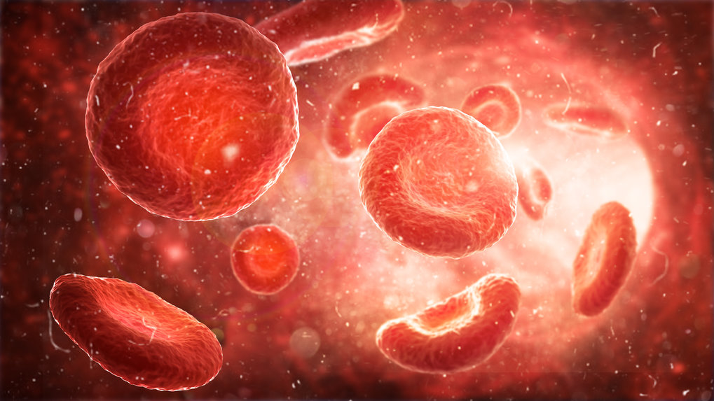
Scientists at the Max Planck Institute of Biochemistry have now discovered why JAK2 mutant tumor cells can survive cancer therapy. Myeloproliferative neoplasms (MPN) is a chronic cancers of the hematopoietic system. In simple terms it is a cancer of blood cells. This can be caused by mutations in JAK2 protein, because this important protein regulates cell division and differentiation. In order to heal the diseases, drugs are needed that prevent the diseased cells from proliferation in the long term. Patients with JAK2 mutations are treated with JAK inhibitors but the treatment often fails. With the currently used drugs, there are some cells that are either resistant to the drugs in advance or get used to the drug. Therefore, these cancer cells continue to divide unintentionally. Scientists around Matthias Mann, from the MPI of Biochemistry together with scientist from the Leibniz Institut on Aging in Jena, have investigated the mechanism that enables cancer cells to survive a drug treatment.

Read more


Publication Placeholder

Bauernfried, S., Scherr, M.J., Pichlmair, A., Duderstadt, K.E., and Hornung, V.
(IMPRS-LS students are in bold)
Science, 2020, Online ahead of print.
doi: 10.1126/science.abd0811

Human NLRP1 is a sensor for double-stranded RNA

Inflammasomes function as intracellular sensors of pathogen infection or cellular perturbation and thereby play a central role in numerous diseases. Given the high abundance of NLRP1 in epithelial barrier tissues, we screened a diverse panel of viruses for inflammasome activation in keratinocytes. We identified Semliki Forest virus (SFV), a positive-strand RNA virus, as a potent activator of human, but not murine NLRP1. SFV replication and the associated formation of double-stranded (ds) RNA was required to engage the NLRP1 inflammasome. Moreover, delivery of long dsRNA was sufficient to trigger activation. Biochemical studies revealed that NLRP1 binds dsRNA via its LRR, resulting in its NACHT domain gaining ATPase activity. Altogether, these results establish human NLRP1 as a direct sensor for dsRNA and thus RNA virus infection.


Publication Placeholder

Karayel, Ö., Xu, P., Bludau, I., Velan Bhoopalan, S., Yao, Y., Ana Rita, F.C., Santos, A., Schulman, B.A., Alpi, A.F., Weiss, M.J., and Mann, M.
Mol Syst Biol 16, e9813.
doi: 10.15252/msb.20209813

Integrative proteomics reveals principles of dynamic phosphosignaling networks in human erythropoiesis

Human erythropoiesis is an exquisitely controlled multistep developmental process, and its dysregulation leads to numerous human diseases. Transcriptome and epigenome studies provided insights into system-wide regulation, but we currently lack a global mechanistic view on the dynamics of proteome and post-translational regulation coordinating erythroid maturation. We established a mass spectrometry (MS)-based proteomics workflow to quantify and dynamically track 7,400 proteins and 27,000 phosphorylation sites of five distinct maturation stages of in vitro reconstituted erythropoiesis of CD34+ HSPCs. Our data reveal developmental regulation through drastic proteome remodeling across stages of erythroid maturation encompassing most protein classes. This includes various orchestrated changes in solute carriers indicating adjustments to altered metabolic requirements. To define the distinct proteome of each maturation stage, we developed a computational deconvolution approach which revealed stage-specific marker proteins. The dynamic phosphoproteomes combined with a kinome-targeted CRISPR/Cas9 screen uncovered coordinated networks of erythropoietic kinases and pinpointed downregulation of c-Kit/MAPK signaling axis as key driver of maturation. Our system-wide view establishes the functional dynamic of complex phosphosignaling networks and regulation through proteome remodeling in erythropoiesis.


Publication Placeholder

Libicher, K., and Mutschler, H.
Chem Commun (Camb), 2020, Online ahead of print.
doi: 10.1039/d0cc06515c

Probing self-regeneration of essential protein factors required for in vitro translation activity by serial transfer

The bottom-up construction of bio-inspired systems capable of self-maintenance and reproduction is a central goal in systems chemistry and synthetic biology. A particular challenge in such systems is the continuous regeneration of key proteins required for macromolecular synthesis. Here, we probe self-maintenance of a reconstituted in vitro translation system challenged by serial transfer of selected key proteins. We find that the system can simultaneously regenerate multiple essential polypeptides, which then contribute to the maintenance of protein expression after serial transfer. The presented strategy offers a robust methodology for probing and optimizing continuous self-regeneration of proteins in cell-free environments.