News

Scientists at the Max Planck Institute (MPI) of Biochemistry enable 100-times faster multiplexed DNA-PAINT microscopy using optimized DNA sequences.

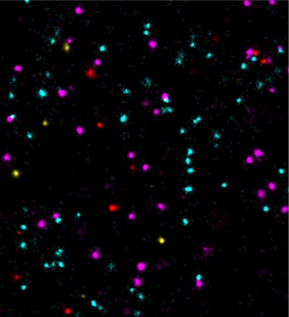
• DNA-PAINT uses DNA-barcoded probes to visualize nanoscale biological structures

• Optimized DNA designs enable 100-times faster and multicolor imaging

• High throughput and molecular resolution microscopy might in the future improve our understanding of the interactions between different tumor markers

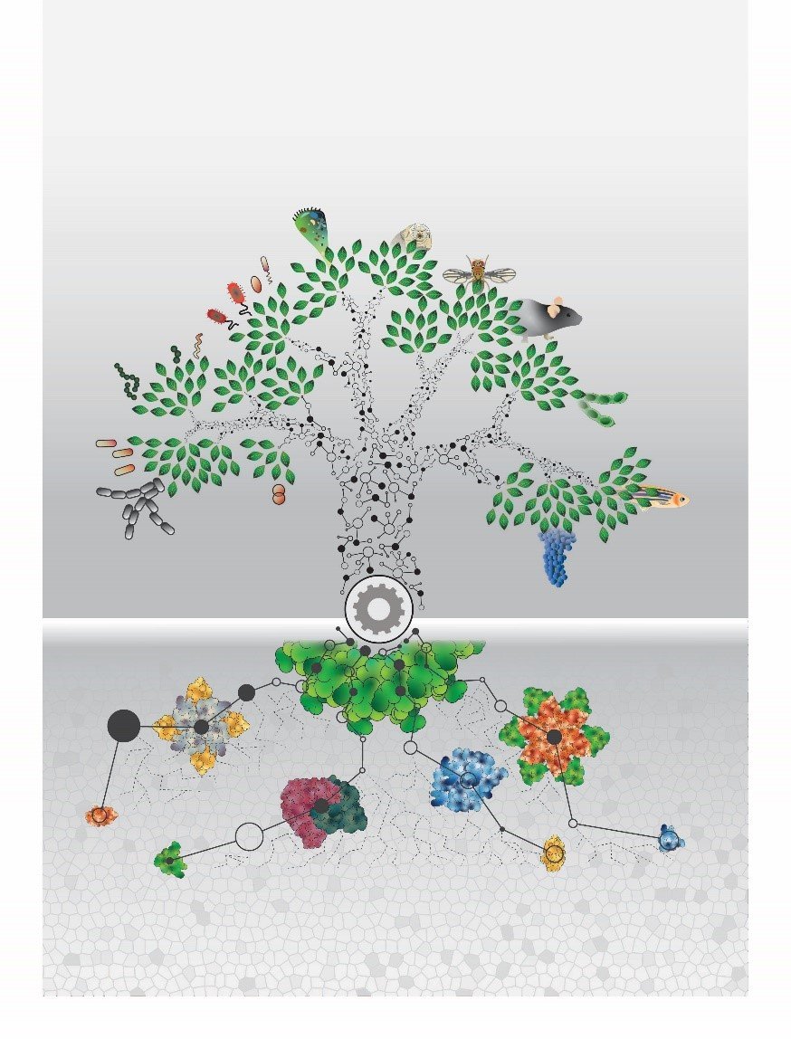
Super-resolution fluorescence microscopy can be used to visualize structures smaller than 200 nanometers, i.e. below the diffraction limit of light. One of the microscopy techniques, called DNA-PAINT, was developed by Ralf Jungmann, research group leader at the MPI of Biochemistry and Professor for Experimental Physics at the Ludwig Maximilian University Munich together with colleagues. The technique uses short ‘imagers’, dye-labeled DNA strands that temporarily bind to their target molecules in a complementary manner to produce the necessary "blinking" for super-resolution reconstruction of the images. “We have recently improved DNA-PAINT’s traditionally rather slow acquisition speed by an order of magnitude by optimizing DNA sequence design.” says Jungmann. "However, this came at the cost of losing multiplexing, which means that several structures in the cell cannot be observed simultaneously", added Jungmann. The simultaneous observation of several proteins, however, is important for the better understanding of complex signaling cascades between tumor and normal cells.”

Read more


Publication Placeholder

Karayel, O., Tonelli, F., Virreira Winter, S., Geyer, P.E., Fan, Y., Sammler, E.M., Alessi, D., Steger, M., and Mann, M.
Mol Cell Proteomics, 2020, [Epub ahead of print].
doi: 10.1074/mcp.RA120.002055

Accurate MS-based Rab10 phosphorylation stoichiometry determination as readout for LRRK2 activity in Parkinson's disease

Pathogenic mutations in the Leucine-rich repeat kinase 2 (LRRK2) are the predominant genetic cause of Parkinson's disease (PD). They increase its activity, resulting in augmented Rab10-Thr73 phosphorylation and conversely, LRRK2 inhibition decreases pRab10 levels. Currently, there is no assay to quantify pRab10 levels for drug target engagement or patient stratification. To meet this challenge, we developed an high accuracy and sensitivity targeted mass spectrometry (MS)-based assay for determining Rab10-Thr73 phosphorylation stoichiometry in human samples. It uses synthetic stable isotope-labeled (SIL) analogues for both phosphorylated and non-phosphorylated tryptic peptides surrounding Rab10-Thr73 to directly derive the percentage of Rab10 phosphorylation from attomole amounts of the endogenous phosphopeptide. The SIL and the endogenous phosphopeptides are separately admitted into an Orbitrap analyzer with the appropriate injection times. We test the reproducibility of our assay by determining Rab10-Thr73 phosphorylation stoichiometry in neutrophils of LRRK2 mutation carriers before and after LRRK2 inhibition. Compared to healthy controls, the PD predisposing mutation carriers LRRK2 G2019S and VPS35 D620N display 1.9-fold and 3.7-fold increased pRab10 levels, respectively. Our generic MS-based assay further establishes the relevance of pRab10 as a prognostic PD marker and is a powerful tool for determining LRRK2 inhibitor efficacy and for stratifying PD patients for LRRK2 inhibitor treatment.


Publication Placeholder

Müller, J.B., Geyer, P.E., Colaço, A.R., Treit, P.V., Strauss, M.T., Oroshi, M., Doll, S., Virreira Winter, S., Bader, J.M., Köhler, N., Theis, F., Santos, A., and Mann, M.
(IMPRS-LS students and -alumni are in bold)
Nature, 2020, 582, 592-596.
doi: 10.1038/s41586-020-2402-x

The proteome landscape of the kingdoms of life

Proteins carry out the vast majority of functions in all biological domains, but for technological reasons their large-scale investigation has lagged behind the study of genomes. Since the first essentially complete eukaryotic proteome was reported, advances in mass-spectrometry-based proteomics have enabled increasingly comprehensive identification and quantification of the human proteome. However, there have been few comparisons across species in stark contrast with genomics initiatives. Here we use an advanced proteomics workflow-in which the peptide separation step is performed by a microstructured and extremely reproducible chromatographic system-for the in-depth study of 100 taxonomically diverse organisms. With two million peptide and 340,000 stringent protein identifications obtained in a standardized manner, we double the number of proteins with solid experimental evidence known to the scientific community. The data also provide a large-scale case study for sequence-based machine learning, as we demonstrate by experimentally confirming the predicted properties of peptides from Bacteroides uniformis. Our results offer a comparative view of the functional organization of organisms across the entire evolutionary range. A remarkably high fraction of the total proteome mass in all kingdoms is dedicated to protein homeostasis and folding, highlighting the biological challenge of maintaining protein structure in all branches of life. Likewise, a universally high fraction is involved in supplying energy resources, although these pathways range from photosynthesis through iron sulfur metabolism to carbohydrate metabolism. Generally, however, proteins and proteomes are remarkably diverse between organisms, and they can readily be explored and functionally compared at www.proteomesoflife.org.


Publication Placeholder

Reim, A., Ackermann, R., Font-Mateu, J., Kammel, R., Beato, M., Nolte, S., Mann, M., Russmann, C., and Wierer, M.
Nat Commun, 2020, 11, 3019.
doi: 10.1038/s41467-020-16837-x

Atomic-resolution mapping of transcription factor-DNA interactions by femtosecond laser crosslinking and mass spectrometry

Transcription factors (TFs) regulate target genes by specific interactions with DNA sequences. Detecting and understanding these interactions at the molecular level is of fundamental importance in biological and clinical contexts. Crosslinking mass spectrometry is a powerful tool to assist the structure prediction of protein complexes but has been limited to the study of protein-protein and protein-RNA interactions. Here, we present a femtosecond laser-induced crosslinking mass spectrometry (fliX-MS) workflow, which allows the mapping of protein-DNA contacts at single nucleotide and up to single amino acid resolution. Applied to recombinant histone octamers, NF1, and TBP in complex with DNA, our method is highly specific for the mapping of DNA binding domains. Identified crosslinks are in close agreement with previous biochemical data on DNA binding and mostly fit known complex structures. Applying fliX-MS to cells identifies several bona fide crosslinks on DNA binding domains, paving the way for future large scale ex vivo experiments.


Publication Placeholder

Lingaraju, M., Schuller, J.M., Falk, S., Gerlach, P., Bonneau, F., Basquin, J., Benda, C., and Conti, E.
Cold Spring Harb Symp Quant Biol, 2020, [Epub ahead of print].
doi: 10.1101/sqb.2019.84.040295

To Process or to Decay: A Mechanistic View of the Nuclear RNA Exosome

The RNA exosome was originally discovered in yeast as an RNA-processing complex required for the maturation of 5.8S ribosomal RNA (rRNA), one of the constituents of the large ribosomal subunit. The exosome is now known in eukaryotes as the major 3'-5' RNA degradation machine involved in numerous processing, turnover, and surveillance pathways, both in the nucleus and the cytoplasm. Yet its role in maturing the 5.8S rRNA in the pre-60S ribosomal particle remains probably the most intricate and emblematic among its functions, as it involves all the RNA unwinding, degradation, and trimming activities embedded in this macromolecular complex. Here, we propose a comprehensive mechanistic model, based on current biochemical and structural data, explaining the dual functions of the nuclear exosome-the constructive versus the destructive mode.


Researchers at the Max Planck Institute of Biochemistry have for the first time uncovered the proteome of 100 organisms from all domains of life. Proteins control life as one of the most important biomolecules - as enzymes, receptors, signal or structural building blocks. Researchers at the Max Planck Institute (MPI) of Biochemistry have for the first time uncovered the proteomes of 100 different organisms. The selected specimens come from all three domains of life: bacteria, archaeae and eukaryotes. Using mass spectrometry, 340,000 unique proteins were measured. Related proteins conserved througout evolution can now be compared quantitatively for the first time. The results were published in Nature.

What do the house mouse Mus Musculus, the organism Haloferax Mediterranei that lives in thermal springs and the intestinal bacterium Escherischia coli have in common? Nothing one would assume, because these three organisms are far distant in their evolutionary ancestry. Each belongs to one of the three different domains, the highest classification category of living organisms: eukaryotes, archaeae or bacteria. All three organisms use similar biomolecules called proteins which are important for their survival. In order to discover new similarities and differences between these and other organisms, researchers from the MPI of Biochemistry, in collaboration with research institutions from Munich and Copenhagen, have analyzed in addition to these three organisms, the proteome of a total of 100 organisms from all domains of life. The proteome is the sum total of the proteins of a cell or living organism. Johannes Müller, one of the two first authors of the study, explains: "Nowadays, evolutionary phylogeny is analysed on the basis of the similarity of certain gene segments. Genes, are the blueprints for proteins. With the current study we have looked at the different gene products, the proteins. We can now determine whether the individual organisms not only carry the building instructions for the proteins, but also produce them.”

Read more


Publication Placeholder

Bader, J.M., Geyer, P.E., Müller, J.B., Strauss, M.T., Koch, M., Leypoldt, F., Koertvelyessy, P., Bittner, D., Schipke, C.G., Incesoy, E.I., Peters, O., Deigendesch, N., Simons, M., Jensen, M.K., Zetterberg, H., and Mann, M.
(IMPRS-LS students and -alumni are in bold)
Mol Syst Biol, 2020, 16, e9356.
doi: 10.15252/msb.20199356

Proteome profiling in cerebrospinal fluid reveals novel biomarkers of Alzheimer's disease

Neurodegenerative diseases are a growing burden, and there is an urgent need for better biomarkers for diagnosis, prognosis, and treatment efficacy. Structural and functional brain alterations are reflected in the protein composition of cerebrospinal fluid (CSF). Alzheimer's disease (AD) patients have higher CSF levels of tau, but we lack knowledge of systems-wide changes of CSF protein levels that accompany AD. Here, we present a highly reproducible mass spectrometry (MS)-based proteomics workflow for the in-depth analysis of CSF from minimal sample amounts. From three independent studies (197 individuals), we characterize differences in proteins by AD status (> 1,000 proteins, CV < 20%). Proteins with previous links to neurodegeneration such as tau, SOD1, and PARK7 differed most strongly by AD status, providing strong positive controls for our approach. CSF proteome changes in Alzheimer's disease prove to be widespread and often correlated with tau concentrations. Our unbiased screen also reveals a consistent glycolytic signature across our cohorts and a recent study. Machine learning suggests clinical utility of this proteomic signature.


Publication Placeholder

Scacchetti, A., Schauer, T., Reim, A., Apostolou, Z., Campos Sparr, A., Krause, S., Heun, P., Wierer, M., and Becker, P.B.
(IMPRS-LS students are in bold)
Elife, 2020, 9.
doi: 10.7554/eLife.56325

Drosophila SWR1 and NuA4 complexes are defined by DOMINO isoforms

Histone acetylation and deposition of H2A.Z variant are integral aspects of active transcription. In Drosophila, the single DOMINO chromatin regulator complex is thought to combine both activities via an unknown mechanism. Here we show that alternative isoforms of the DOMINO nucleosome remodeling ATPase, DOM-A and DOM-B, directly specify two distinct multi-subunit complexes. Both complexes are necessary for transcriptional regulation but through different mechanisms. The DOM-B complex incorporates H2A.V (the fly ortholog of H2A.Z) genome-wide in an ATP-dependent manner, like the yeast SWR1 complex. The DOM-A complex, instead, functions as an ATP-independent histone acetyltransferase complex similar to the yeast NuA4, targeting lysine 12 of histone H4. Our work provides an instructive example of how different evolutionary strategies lead to similar functional separation. In yeast and humans, nucleosome remodeling and histone acetyltransferase complexes originate from gene duplication and paralog specification. Drosophila generates the same diversity by alternative splicing of a single gene.


Publication Placeholder

Soni, K., Martínez-Lumbreras, S., and Sattler, M.
J Mol Biol, 2020, [Epub ahead of print].
doi: 10.1016/j.jmb.2020.05.012

Conformational dynamics from ambiguous zinc coordination in the RanBP2-type zinc finger of RBM5

The multi-domain RNA binding protein RBM5 is a molecular signature of metastasis. RBM5 regulates alternative splicing of apoptotic genes including the cell death receptor Fas and the initiator Caspase-2. The RBM5 RanBP2-type zinc finger (Zf1) is known to specifically recognize single stranded RNAs with high affinity. Here, we study the structure and conformational dynamics of the Zf1 zinc finger of human RBM5 using NMR. We show that the presence of a non-canonical cysteine in Zf1 kinetically destabilizes the protein. Metal exchange kinetics show that mutation of the cysteine establishes high affinity coordination of the zinc. Our data indicate that selection of such a structurally destabilizing mutation during the course of evolution could present an opportunity for functional adaptation of the protein.


Publication Placeholder

Schmacke, N.A., and Hornung, V.
Nature, 2020, 581, 266-267.
doi: 10.1038/d41586-020-0133


Fourth defence molecule completes antiviral line-up

Toll-like receptors can initiate an immune response when they detect signs of a viral or microbial threat. New insight into how such receptor activation drives defence programs should aid our efforts to understand autoimmune diseases.