News

Yunmin Wu is interested in how we perceive motion. Inspired by a cat video, she came up with the elegant idea to elicit the waterfall illusion in tiny zebrafish larvae. Thereby, the PhD graduate in Fumi Kubo’s1 group from the department of Herwig Baier gained surprising insights into the neuronal mechanism of seeing motion.

Can you elaborate on your topic of research and why you work with the model organism zebrafish?

Inside the brain, there are many neurons that process motion and its direction. As these neurons are quite abundant, I am interested to find out if all of them are required to recognize motion. For this, larval zebrafish is an amazing animal model – it is small, transparent, and demonstrates so many complex behaviors that we humans also display.

How did you come up with the idea of an optical illusion as a tool to study motion processing?

I was inspired by a video, in which a cat was trying to capture an illusory moving snake. I asked myself if an illusion that affects us humans might also apply to zebrafish. If that is the case, I can then look into the brain and see which neurons are involved. With the help of my supervisor Fumi, I chose the motion aftereffect eventually among many other cool illusions.

Read more


graduationCongratulations on your PhD!


Kira Bartnik
Single-Molecule FRET Studies of Protein Function and Conformational Dynamics -
From DNA Nanotechnology to Viral and Bacterial Infections
RG: Don Lamb


Publication Placeholder

Wu, Y., Dal Maschio, M., Kubo, F., and Baier, H.
Neuron, 2020, [Epub ahead of print].
doi: 10.1016/j.neuron.2020.08.027

An Optical Illusion Pinpoints an Essential Circuit Node for Global Motion Processing

Direction-selective (DS) neurons compute the direction of motion in a visual scene. Brain-wide imaging in larval zebrafish has revealed hundreds of DS neurons scattered throughout the brain. However, the exact population that causally drives motion-dependent behaviors-e.g., compensatory eye and body movements-remains largely unknown. To identify the behaviorally relevant population of DS neurons, here we employ the motion aftereffect (MAE), which causes the well-known "waterfall illusion." Together with region-specific optogenetic manipulations and cellular-resolution functional imaging, we found that MAE-responsive neurons represent merely a fraction of the entire population of DS cells in larval zebrafish. They are spatially clustered in a nucleus in the ventral lateral pretectal area and are necessary and sufficient to steer the entire cycle of optokinetic eye movements. Thus, our illusion-based behavioral paradigm, combined with optical imaging and optogenetics, identified key circuit elements of global motion processing in the vertebrate brain.


Publication Placeholder

Tüshaus, J., Müller, S.A., Kataka, E.S., Zaucha, J., Sebastian Monasor, L., Su, M., Güner, G., Jocher, G., Tahirovic, S., Frishman, D., Simons, M., and Lichtenthaler, S.F.
(IMPRS_LS students are in bold)
EMBO J, 2020, e105693, online ahead of print.
doi: 10.15252/embj.2020105693

An optimized quantitative proteomics method establishes the cell type-resolved mouse brain secretome

To understand how cells communicate in the nervous system, it is essential to define their secretome, which is challenging for primary cells because of large cell numbers being required. Here, we miniaturized secretome analysis by developing the "high-performance secretome protein enrichment with click sugars" (hiSPECS) method. To demonstrate its broad utility, hiSPECS was used to identify the secretory response of brain slices upon LPS-induced neuroinflammation and to establish the cell type-resolved mouse brain secretome resource using primary astrocytes, microglia, neurons, and oligodendrocytes. This resource allowed mapping the cellular origin of CSF proteins and revealed that an unexpectedly high number of secreted proteins in vitro and in vivo are proteolytically cleaved membrane protein ectodomains. Two examples are neuronally secreted ADAM22 and CD200, which we identified as substrates of the Alzheimer-linked protease BACE1. hiSPECS and the brain secretome resource can be widely exploited to systematically study protein secretion and brain function and to identify cell type-specific biomarkers for CNS diseases.


Publication Placeholder

Agarwal, R., and Duderstadt, K.E.
Nat Commun, 2020, 11, 4714.
doi: 10.1038/s41467-020-18456-y

Multiplex flow magnetic tweezers reveal rare enzymatic events with single molecule precision

The application of forces and torques on the single molecule level has transformed our understanding of the dynamic properties of biomolecules, but rare intermediates have remained difficult to characterize due to limited throughput. Here, we describe a method that provides a 100-fold improvement in the throughput of force spectroscopy measurements with topological control, which enables routine imaging of 50,000 single molecules and a 100 million reaction cycles in parallel. This improvement enables detection of rare events in the life cycle of the cell. As a demonstration, we characterize the supercoiling dynamics and drug-induced DNA break intermediates of topoisomerases. To rapidly quantify distinct classes of dynamic behaviors and rare events, we developed a software platform with an automated feature classification pipeline. The method and software can be readily adapted for studies of a broad range of complex, multistep enzymatic pathways in which rare intermediates have escaped classification due to limited throughput.


Publication Placeholder

Gehrlach, D.A., Weiand, C., Gaitanos, T.N., Cho, E., Klein, A.S., Hennrich, A.A., Conzelmann, K.K., and Gogolla, N. (IMPRS-LS students are in bold)
Elife, 2020, 9.
doi: 10.7554/eLife.55585

A whole-brain connectivity map of mouse insular cortex

The insular cortex (IC) plays key roles in emotional and regulatory brain functions and is affected across psychiatric diseases. However, the brain-wide connections of the mouse IC have not been comprehensively mapped. Here, we traced the whole-brain inputs and outputs of the mouse IC across its rostro-caudal extent. We employed cell-type-specific monosynaptic rabies virus tracings to characterize afferent connections onto either excitatory or inhibitory IC neurons, and adeno-associated viral tracings to label excitatory efferent axons. While the connectivity between the IC and other cortical regions was highly bidirectional, the IC connectivity with subcortical structures was often unidirectional, revealing prominent cortical-to-subcortical or subcortical-to-cortical pathways. The posterior and medial IC exhibited resembling connectivity patterns, while the anterior IC connectivity was distinct, suggesting two major functional compartments. Our results provide insights into the anatomical architecture of the mouse IC and thus a structural basis to guide investigations into its complex functions.


Support for the new Max Planck life science campus in Martinsried and the initiative for quantum computing and quantum technologies

Mastering transformation with technology – that is the motto of the Bavarian State Government, whose Hightech Agenda saw Bavaria launch Germany’s only technology offensive of its kind a year ago. With an injection of EUR 2 billion, 1,000 new professorships and 13,000 new university places, the Free State of Bavaria wants to build upon its leading position in research. The Max-Planck-Gesellschaft welcomes the fact that, with the latest cabinet decision of 14 September 2020, significant additional funding is being committed to cutting-edge research, including at Max Planck Institutes. At a press conference, Bavarian Prime Minister Markus Söder raised the prospect of significant support for the establishment of a life science campus in Martinsried for interdisciplinary and interactive research in the biosciences. On the Martinsried campus, the Max-Planck-Gesellschaft plans to establish a new Max Planck Institute for the Study of Life with a view to pooling and realigning its existing strengths. With an equivalent of 18 Departments, the Institute will be the largest of the Max Planck Institutes and will initially focus on the synthesis of artificial cells and the study of the brain in its natural environment (real-life neuroscience).

Read more


Peter Krenn from the Department Molecular Medicine (Director Reinhard Fässler) at the Max Planck Institute of Biochemistry has found a new way to attack leukemic stem cells.

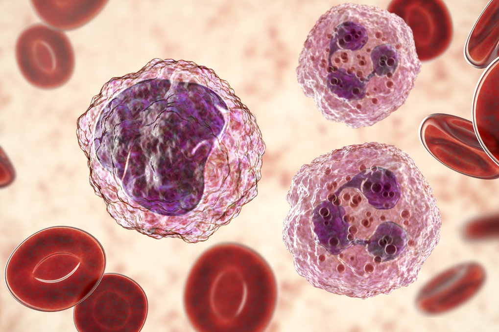
Blood – the juice of life Blood supplies complex organisms with nutritive substances, transports metabolic products or messenger substances. Cellular components of blood are erythrocytes responsible for the transport of oxygen and carbon dioxide, thrombocytes responsible for blood coagulation and leukocytes responsible for the immune defense. As each of these blood cells has a limited lifespan, and are massively lost during bleeding or consumed during infections. Hence, they must be continuously replaced, which is ensured by the so-called hematopoietic stem cells in the bone marrow. These cells have the ability to develop into any type of blood cell. Chronic myeloid leukemia In chronic myeloid leukemia, the hematopoietic stem cell undergoes a genetic mutation by recombining chromosome 9 and 22. As a result, gene building blocks fuse that would otherwise not be in contact with each other. The incorrectly assembled chromosome is called Philadelphia chromosome and harbors the construction manual for the so-called BCR-ABL oncogene. This causes the leukemic stem cell to behave selfishly: it divides and multiplies drastically at the expense of healthy blood stem cells.

Read more


graduationCongratulations on your PhD!


Daniel Gehrlach
Anatomical and functional characterization of the mouse insular cortex
RG: Nadine Gogolla

Daniil Markov
Involvement of cerebellar Purkinje cells in adaptive locomotion of larval zebrafish
RG: Ruben Portugues


MPIB scientists have contributed to deciphering the 3D structure of the nuclear pore of baker's yeast cells.

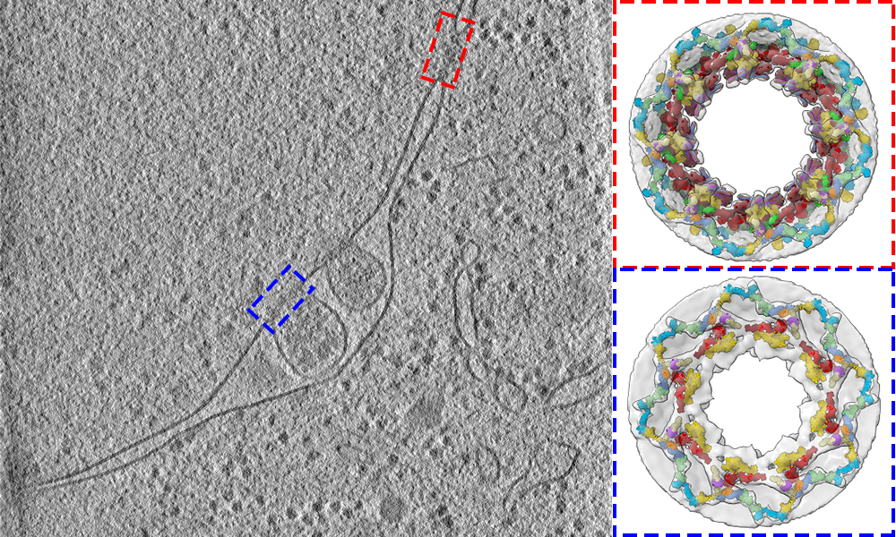
Research group leader Boris Pfander and his team from the Max Planck Institute of Biochemistry, together with colleagues from the Max Planck Institute of Biophysics in Frankfurt am Main and the EMBL in Heidelberg, have investigated the 3D structure of nuclear pores in budding yeast (Saccharomyces cerevisiae). Their results, published in Nature, reveal the architecture of the nuclear pore complex in intact cells and broaden our understanding of crucial processes in life.

Nuclear pores are a highly complex assembly of proteins. Hundreds of them are embedded in the double membrane that surrounds and protects the cell’s nucleus. They act as a gateway that regulates the entry and exit of molecules. An important function of nuclear pores is to regulate the export of a molecule called messenger RNA (mRNA) from the nucleus into the surrounding cell – the cytoplasm – where it delivers instructions for the assembly of proteins. Revealing the architecture Now the scientists appreciate better how the nuclear pore works in its native context, how it is maintained and recycled. The study provides a detailed structural description of the three protein rings that make up the nuclear pore, known as the cytoplasmic, nuclear, and inner rings. To show how these rings are arranged in cells, the researchers used a combination of cell biology, computational modelling, and in-cell cryo-electron tomography: an imaging technique, that is used to produce high-resolution 3D views of the molecular landscape inside a cell. This led to fundamental new insights. The scientist found out that the 3D configuration of the cytoplasmic ring accommodates the path of mRNA export.

Read more